


Page 13 - Read Online
P. 13
Page 8 of 14                                                      Lust et al. Vessel Plus 2021;5:7  I  http://dx.doi.org/10.20517/2574-1209.2020.52


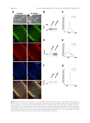
               Figure 2. Pressure stimulation of isolated mouse veins. Isolated branches of the mesenteric vein of male NMRI mice were exposed to
               pressure levels of 4 or 16 mmHg, respectively. Afterwards, vessel segments were processed for whole mount immunofluorescence
               staining, scale bars: 0.1 mm (A) to detect CD31 (endothelial cell marker), αSMA (smooth muscle cell marker), and DAPI (visualization
               of the nuclei). Protein samples generated from these blood vessels were pooled and analyzed by automated capillary electrophoresis/
               immunodetection [required due to the low yield of protein; specified antigens were detected by antibodies and corresponding signals
               (B, D, F) and were automatically evaluated by their size (kDa) and intensity (area under the curve, C, E, G)]. Signals specific for COX-1 (B
               and C) and COX-2 (D and E) were detected. b-actin served as a loading reference (F and G).

