


Page 69 - Read Online
P. 69
Barioli et al. Vessel Plus 2024;8:13  https://dx.doi.org/10.20517/2574-1209.2023.68   Page 7 of 14


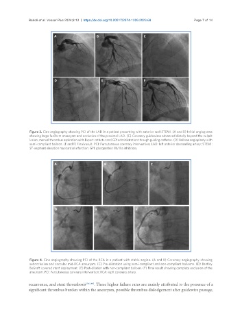
                Figure 3. Cine angiography showing PCI of the LAD in a patient presenting with anterior wall STEMI. (A and B) Initial angiograms
                showing large fusiform aneurysm and occlusion of the proximal LAD. (C) Coronary guidewires advanced distally beyond the culprit
                lesion, manual thrombus aspiration with Export catheter and GPI administration through guiding catheter. (D) Balloon angioplasty with
                semi-compliant balloon. (E and F) Final result. PCI: Percutaneous coronary intervention; LAD: left anterior descending artery; STEMI:
                ST-segment elevation myocardial infarction. GPI: glycoprotein IIb/IIIa inhibitors.


                Figure 4. Cine angiography showing PCI of the RCA in a patient with stable angina. (A and B) Coronary angiography showing
                subocclusion and saccular mid-RCA aneurysm. (C) Pre-dilatation using semi-compliant and non-compliant balloons. (D) Bentley
                BeGraft covered stent deployment. (E) Post-dilation with non-compliant balloon. (F) Final result showing complete exclusion of the
                aneurysm. PCI: Percutaneous coronary intervention; RCA: right coronary artery.

               recurrence, and stent thrombosis [5,43,44] . These higher failure rates are mainly attributed to the presence of a
               significant thrombus burden within the aneurysm, possible thrombus dislodgement after guidewire passage,

