


Page 67 - Read Online
P. 67
Barioli et al. Vessel Plus 2024;8:13  https://dx.doi.org/10.20517/2574-1209.2023.68   Page 5 of 14

               Table 1. Therapeutic strategies for CAAs and CAEs
                Treatment modality    Rationale             Advantages           Disadvantages
                Medical   ACEi/ARBs/Statins  Control CVRF/Slow CAA dilation  Easy access   Neither removal nor exclusion of CAA
                therapy               rate?                 Wide availability    Risk of bleeding with long-term
                                                                                 antithrombotic therapies
                       DAPT vs. SAPT vs.   Reduce thrombotic
                       OAC            complications
                       IVIG therapy   Kawasaki disease-related CAA
                PCI    Covered Stent  Exclusion of smaller CAA   Lower risk or procedural   No removal of CAA/risk of CAA
                                      Prevention of CAA rupture   complications/quick coronary   rupture/endoleaks/stent
                       Double stent method
                                      Coronary flow restoration in case  flow restoration  malapposition/vessel occlusion
                       Self-expanding stents  of ongoing ischemia
                       Coil embolization ±
                       stent-assisted
                Cardiac   Resection/ligation/   Treatment of larger/giant CAA   Complete removal of CAA   Higher risk of procedural complications
                surgery  marsupialization  at high risk of rupture or   Treatment of CAA/CAD
                                      involving LMCA/proximal vessel  unsuitable for PCI
                       CABG           Treatment of concomitant
                                      obstructive CAD
               ACEi: Angiotensin converting enzyme inhibitors; ARBs: angiotensin II receptor blockers; DAPT: dual antiplatelet therapy; SAPT: single antiplatelet
               therapy; OAC: oral anticoagulant; CAA: coronary artery aneurysm; CAE: coronary artery ectasia; CVRF: cardiovascular risk factors; PCI:
               percutaneous coronary intervention; CABG: coronary artery bypass graft; LMCA: left main coronary artery; CAD: coronary artery disease.


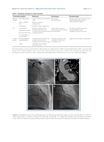
                Figure 2. Angiographic (A) and CT-scan (B) appearance of a LAD saccular aneurysm (double arrows) in a patient with prior CABG. (C
                and D) show patency of LIMA to LAD and SVG to OM grafts, respectively. CT: Computed tomography; LAD: left anterior descending
                artery; CABG: coronary artery bypass grafting; LIMA: left internal mammary artery; SVG: saphenous vein graft; OM: obtuse marginal.

