


Page 9 - Read Online
P. 9
Page 6 of 39                           Yang et al. Soft Sci. 2025, 5, 46  https://dx.doi.org/10.20517/ss.2025.44


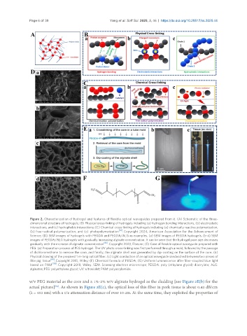
                Figure 2. Characterization of hydrogel and features of flexible optical waveguides prepared from it. (A) Schematic of the three-
                dimensional structure of hydrogels; (B) Physical cross-linking of hydrogels including (a) hydrogen bonding interactions, (b) electrostatic
                interactions, and (c) hydrophobic interactions; (C) Chemical cross-linking of hydrogels including (a) chemically reactive polymerization,
                (b) free-radical polymerization, and (c)  photopolymerization [56] . Copyright 2024, American Association for the Advancement of
                Science; (D) SEM images of hydrogels with PEGDA and PEGDA/ALG as examples. (a) SEM images of PEGDA hydrogels. (b-d) SEM
                images of PEGDA/ALG hydrogels with gradually increasing alginate concentration. It can be seen that the hydrogel pore size decreases
                                                   [86]
                gradually with the increase of alginate concentration  . Copyright 2023, Elsevier; (E) Case of flexible optical waveguide prepared with
                PEG. (a) Preparation process of PEG hydrogel. The UV photo cross-linking was first performed through a mold, followed by the passage
                of dichloromethane to remove the core, and finally, the alginate shell was generated by dip-coating on the surface of the core. (b)
                Physical drawing of the prepared 1 m-long optical fiber. (c) Light conduction of an optical waveguide sandwiched between two pieces of
                         [94]
                thin pig  tissue  . Copyright 2015, Wiley; (F) Chemical formula of PEGDA; (G) Uniform luminescence after fiber-coupled blue light
                          [105]
                based on  PAM  . Copyright 2018, Wiley. SEM: Scanning electron microscope; PEGDA: poly (ethylene glycol) diacrylate; ALG:
                alginates; PEG: polyethylene glycol; UV: ultraviolet; PAM: polyacrylamide.
               w/v PEG material as the core and a 1%-2% w/v alginate hydrogel as the cladding [see Figure 2E(b) for the
                           [94]
               actual picture] . As shown in Figure 2E(c), the optical loss of this fiber in pork tissue is about 0.42 dB/cm
               (λ = 402 nm) with a 1/e attenuation distance of over 10 cm. At the same time, they exploited the properties of

