
Page 95 - Read Online
P. 95
Kim et al. Soft Sci 2024;4:24 https://dx.doi.org/10.20517/ss.2024.09 Page 21 of 27
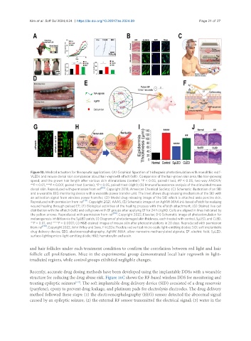
Figure 10. Medical actuators for therapeutic applications. (A) Graphical figuration of trichogenic photostimulation with monolithic red f-
VLEDs and mouse dorsal skin comparison about hair-regrowth effect (left). Comparison of the hair-grown skin area, the hair-growing
speed, and the grown hair length after various skin stimulations (center). *P < 0.05, paired t test, #P < 0.05, two way ANOVA;
**P < 0.01, ***P < 0.001, paired t test (center), *P < 0.05, paired t test (right); (B) Immunofluorescence analysis of the stimulated mouse
dorsal skin. Reproduced with permission from ref [116] . Copyright 2018, American Chemical Society; (C) Schematic illustration of an SID
and a wearable EEG monitoring device with a wearable power transfer unit. The inset shows drug releasing mechanism of the SID with
an activation signal from wireless power transfer; (D) Model drug releasing image of the SID which is attached onto porcine skin.
Reproduced with permission from ref [117] . Copyright 2021, AAAS; (E) Schematic image of an AgNW-MAA ink-based ePatch for realizing
wound healing through pulsed EF; (F) Biological activities of the healing process with the ePatch attachment; (G) Stained live cell
distribution with the ePatch (left) and cell grown with EF groups after applying EF for 24 h (right). Cells are aligned in lines indicated by
the yellow arrows. Reproduced with permission from ref [118] . Copyright 2022, Elsevier; (H) Schematic image of photostimulation for
melanogenesis inhibition via the SµLED patch; (I) Diagram of photodamaged skin thickness, each treated with control, SµLED, and CLED.
**P = 0.01, and ****P = 0.0001; (J) H&E-stained images of mouse skin after photostimulations in 20 days. Reproduced with permission
from ref [119] . Copyright 2022, John Wiley and Sons. f-VLEDs: Flexible red vertical micro-scale light-emitting diodes; SID: soft implantable
drug delivery device; EEG: electroencephalography; AgNW-MAA: silver nanowire-methacrylated alginate; EF: electric field; SµLED:
surface-lighting micro-light-emitting diode; H&E: hematoxylin and eosin.
and hair follicles under each treatment condition to confirm the correlation between red light and hair
follicle cell proliferation. Mice in the experimental group demonstrated local hair regrowth in light-
irradiated regions, while control groups exhibited negligible changes.
Recently, accurate drug dosing methods have been developed using the implantable DDSs with a wearable
structure for reducing the drug abuse risk. Figure 10C shows the RF-based wireless DDS for monitoring and
treating epileptic seizures . The soft implantable drug delivery device (SID) consisted of a drug reservoir
[117]
(parylene), epoxy to prevent drug leakage, and platinum pads for electrolysis electrodes. The drug delivery
method followed these steps: (1) the electroencephalography (EEG) sensor detected the abnormal signal
caused by an epileptic seizure; (2) the external RF sensor transmitted the electrical signal; (3) water in the