
Page 89 - Read Online
P. 89
Yang et al. Plast Aesthet Res 2020;7:8 I http://dx.doi.org/10.20517/2347-9264.2019.63 Page 5 of 13
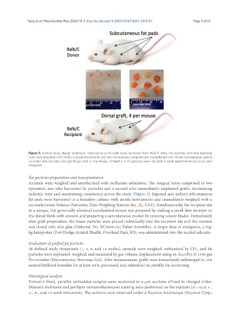
Figure 3. Animal study design schematic. Subcutaneous fat pads were harvested from Balb/C mice. Fat particles with two diameter
sizes were prepared with sterile surgical instruments and then immediately syngeneically-transplanted into dorsal subcutaneous spaces
via small skin incisions, two points per side in one mouse. At least n = 10 particles were included in each experimental group at each
timepoint
Fat particles preparation and transplantation
Animals were weighed and anesthetized with isoflurane inhalation. The surgical team comprised of two
operators, one who harvested fat particles and a second who immediately implanted grafts, minimizing
ischemic time and maintaining consistency across the study [Figure 3]. Inguinal and axillary subcutaneous
fat pads were harvested in a biosafety cabinet with sterile instruments and immediately weighed with a
microelectronic balance (Sartorius, Data Weighing Systems Inc., IL, USA). Simultaneously, the recipient site
in a unique, but genetically identical anesthetized mouse was prepared by making a small skin incision on
the dorsal flank with scissors and preparing a subcutaneous pocket by opening scissor blades. Immediately
after graft preparation, the tissue particles were placed individually into the recipient site and the incision
was closed with skin glue (Vetbond, No. NC9604126; Fisher Scientific). A single dose of analgesics, 5 mg/
kg ketoprofen (Fort Dodge Animal Health, Overland Park, KS), was administered into the nuchal subcutis.
Evaluation of grafted fat particles
At defined study timepoints (1, 4, 8, and 12 weeks), animals were weighed, euthanized by CO , and fat
2
particles were explanted, weighed, and measured by gas volume displacement using an AccuPyc II 1340 gas
Pycnometer (Micrometrics, Norcross, GA). After measurement, grafts were immediately submerged in 10%
neutral buffered formalin for at least 24 h, processed, and embedded in paraffin for sectioning.
Histological analysis
Formalin fixed, paraffin embedded samples were sectioned to 6-μm sections affixed to charged slides.
Masson’s trichrome and perilipin immunofluorescent staining were performed on the explants (n = 6) at 1-,
4-, 8-, and 12-week timepoints. The sections were observed under a Keyence microscope (Keyence Corp.,