
Page 87 - Read Online
P. 87
Yang et al. Plast Aesthet Res 2020;7:8 I http://dx.doi.org/10.20517/2347-9264.2019.63 Page 3 of 13
A
B
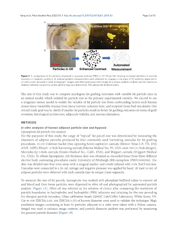
Figure 1. A: comparison of fat particles dispersed in aqueous solution (PBS) or Oil (Olive Oil) showing increased definition of particle
boundary in lipophilic solutions; B: adipose particle measurements were obtained by imaging a minimum of 50 particles dispersed in
oil with a ruler included in each photograph. Images were then processed with ImageJ to increase particle contrast and the maximum
distance between the particle center and the edge was determined. PBS: phosphate-buffered saline
The aim of this study was to compare autologous fat grafting outcomes with variable fat particle sizes in
an animal model, which isolated fat particle size as the primary experimental variable. We elected to use
a syngeneic mouse model to isolate the variable of fat particle size from confounding factors such human
donor tissue variability, trauma from tissue harvest, ischemic time, and recipient tissue bed vascularity. Our
overall study goal was to clarify if smaller fat particles result in better fat grafting outcomes in terms of graft
retention, histological architecture, adipocyte viability, and neovascularization.
METHODS
In vitro analysis of human adipose particle size and hypoxia
Lipoaspirate fat particle size analysis
For the purposes of this study, the range of “typical” fat parcel size was determined by measuring the
diameters of adipose particles produced by four commonly used harvesting cannulas for fat grafting
procedures: 15-cm Coleman-bucket (two opposing holes) aspiration cannula (Mentor Texas L.P., TX, USA
#COL-ASPI), Khouri 12-hole harvesting cannula [Marina Medical Inc. FL, USA; #800-205 (12-hole design)],
Mercedes tip 3-hole cannula (Grams Medical Inc., Calif., USA), and Shippert cannula (Shippert Medical
Co., USA). To obtain lipoaspirate, full thickness skin was obtained as discarded tissue from three different
elective body contouring procedures under University of Pittsburgh IRB exemption (PRO13090506). The
skin was divided into four even areas with a surgical marker and evenly infused with 0.9% NaCl solution.
Cannulas were connected to a 20-mL syringe and negative pressure was applied by hand. At least 10 mL of
adipose particles were obtained with each cannula type in unique tissue segments.
To measure the size of fat parcels, lipoaspirate was washed with phosphate buffered saline to remove oil
and blood and then tissue particles were dispersed in olive oil and photographed for automated particle
analysis [Figure 1A]. Olive oil was selected as the solution of choice after comparing the resolution of
particle boundaries in hydrophobic and hydrophilic (PBS) solutions and selecting for the one providing
the sharpest particle boundary. Glass calibration beads (QAQC Lab/Coffee Laboratory, White Stone, VA.
Cat #s 600 ZSICSA-2.00, 600 ZSICSA-3.35) of known diameter were used to validate the technique. High
resolution images containing at least 50 particles adjacent to a ruler were taken with a Nikon camera.
ImageJ was used to enhance image contrast, and particle diameter analysis was performed by measuring
the greatest particle diameter [Figure 1B].