
Page 88 - Read Online
P. 88
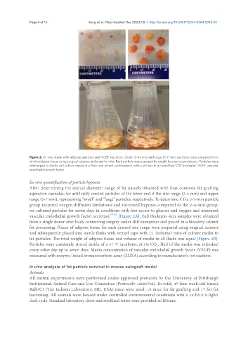
Page 4 of 13                                              Yang et al. Plast Aesthet Res 2020;7:8  I  http://dx.doi.org/10.20517/2347-9264.2019.63


                                        A


                                        B


               Figure 2. Ex vivo study with adipose particles and VEGF secretion. Small (2-4 mm) and large (5-7 mm) particles were prepared from
               whole adipose tissue using surgical scissors and a sterile ruler. Each particle was assessed by weight to ensure consistency. Particles were
               submerged in sterile cell culture media in a flask and stirred continuously with a stir bar in a humidified CO2 incubator. VEGF: vascular
               endothelial growth factor


               Ex vivo quantification of particle hypoxia
               After determining the typical diameter range of fat parcels obtained with four common fat grafting
               aspiration cannulas, we artificially created particles of the lower end if the size range (2-4 mm) and upper
               range (5-7 mm), representing “small” and “large” particles, respectively. To determine if the 5-7-mm particle
               group incurred oxygen diffusion limitations and increased hypoxia compared to the 2-4-mm group,
               we cultured particles for seven days in conditions with free access to glucose and oxygen and measured
               vascular endothelial growth factor secretion [18,19]  [Figure 2A]. Full thickness skin samples were obtained
               from a single donor after body contouring surgery under IRB exemption and placed in a biosafety cabinet
               for processing. Pieces of adipose tissue for each desired size range were prepared using surgical scissors
               and subsequently placed into sterile flasks with vented caps with 1:1 (volume) ratio of culture media to
               fat particles. The total weight of adipose tissue and volume of media in all flasks was equal [Figure 2B].
               Particles were constantly stirred inside of a 37 °C incubator, at 5% CO . Half of the media was refreshed
                                                                            2
               every other day up to seven days. Media concentration of vascular endothelial growth factor (VEGF) was
               measured with enzyme-linked immunosorbent assay (ELISA) according to manufacturer’s instructions.

               In vivo  analysis of fat particle survival in mouse autograft model
               Animals
               All animal experiments were performed under approved protocols by the University of Pittsburgh
               Institutional Animal Care and Use Committee (Protocol# 12080782). In total, 47 four-week-old female
               Balb/CJ (The Jackson Laboratory, ME, USA) mice were used: 18 mice for fat grafting and 17 for fat
               harvesting. All animals were housed under controlled environmental conditions with a 12-h/12-h light/
               dark cycle. Standard laboratory chow and sterilized water were provided ad libitum.