
Page 144 - Read Online
P. 144
Lin et al. Plast Aesthet Res 2019;6:16 I http://dx.doi.org/10.20517/2347-9264.2019.23 Page 7 of 10
A B
C D
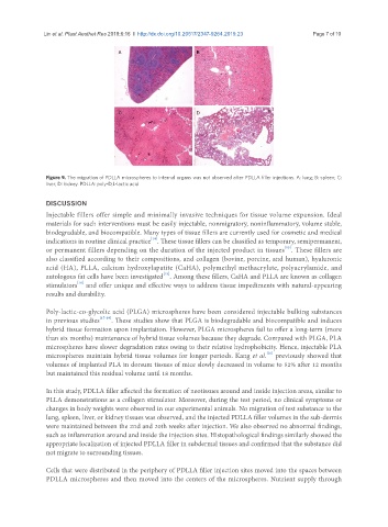
Figure 9. The migration of PDLLA microspheres to internal organs was not observed after PDLLA filler injections. A: lung; B: spleen; C:
liver; D: kidney. PDLLA: poly-D,L-lactic acid
DISCUSSION
Injectable fillers offer simple and minimally invasive techniques for tissue volume expansion. Ideal
materials for such interventions must be easily injectable, nonmigratory, noninflammatory, volume stable,
biodegradable, and biocompatible. Many types of tissue fillers are currently used for cosmetic and medical
[14]
indications in routine clinical practice . These tissue fillers can be classified as temporary, semipermanent,
[15]
or permanent fillers depending on the duration of the injected product in tissues . These fillers are
also classified according to their compositions, and collagen (bovine, porcine, and human), hyaluronic
acid (HA), PLLA, calcium hydroxylapatite (CaHA), polymethyl methacrylate, polyacrylamide, and
[15]
autologous fat cells have been investigated . Among these fillers, CaHA and PLLA are known as collagen
[16]
stimulators and offer unique and effective ways to address tissue impediments with natural-appearing
results and durability.
Poly-lactic-co-glycolic acid (PLGA) microspheres have been considered injectable bulking substances
in previous studies [17-19] . These studies show that PLGA is biodegradable and biocompatible and induces
hybrid tissue formation upon implantation. However, PLGA microspheres fail to offer a long-term (more
than six months) maintenance of hybrid tissue volumes because they degrade. Compared with PLGA, PLA
microspheres have slower degradation rates owing to their relative hydrophobicity. Hence, injectable PLA
[20]
microspheres maintain hybrid tissue volumes for longer periods. Kang et al. previously showed that
volumes of implanted PLA in dorsum tissues of mice slowly decreased in volume to 52% after 12 months
but maintained this residual volume until 18 months.
In this study, PDLLA filler affected the formation of neotissues around and inside injection areas, similar to
PLLA demonstrations as a collagen stimulator. Moreover, during the test period, no clinical symptoms or
changes in body weights were observed in our experimental animals. No migration of test substance to the
lung, spleen, liver, or kidney tissues was observed, and the injected PDLLA filler volumes in the sub-dermis
were maintained between the 2nd and 20th weeks after injection. We also observed no abnormal findings,
such as inflammation around and inside the injection sites. Histopathological findings similarly showed the
appropriate localization of injected PDLLA filler in subdermal tissues and confirmed that the substance did
not migrate to surrounding tissues.
Cells that were distributed in the periphery of PDLLA filler injection sites moved into the spaces between
PDLLA microspheres and then moved into the centers of the microspheres. Nutrient supply through