
Page 141 - Read Online
P. 141
Page 4 of 10 Lin et al. Plast Aesthet Res 2019;6:16 I http://dx.doi.org/10.20517/2347-9264.2019.23
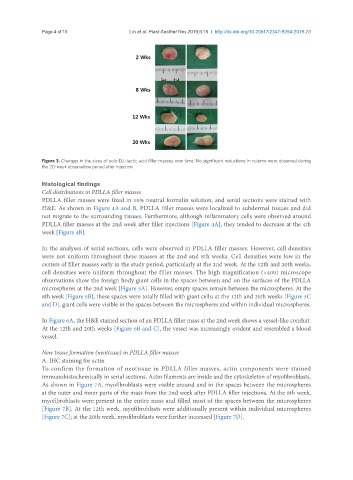
Figure 3. Changes in the sizes of poly-D,L-lactic acid filler masses over time. No significant reductions in volume were observed during
the 20-week observation period after injection
Histological findings
Cell distributions in PDLLA filler masses
PDLLA filler masses were fixed in 10% neutral formalin solution, and serial sections were stained with
H&E. As shown in Figure 4A and B, PDLLA filler masses were localized to subdermal tissues and did
not migrate to the surrounding tissues. Furthermore, although inflammatory cells were observed around
PDLLA filler masses at the 2nd week after filler injections [Figure 4A], they tended to decrease at the 4th
week [Figure 4B].
In the analyses of serial sections, cells were observed in PDLLA filler masses. However, cell densities
were not uniform throughout these masses at the 2nd and 8th weeks. Cell densities were low in the
centers of filler masses early in the study period, particularly at the 2nd week. At the 12th and 20th weeks,
cell densities were uniform throughout the filler masses. The high magnification (×400) microscope
observations show the foreign body giant cells in the spaces between and on the surfaces of the PDLLA
microspheres at the 2nd week [Figure 5A]. However, empty spaces remain between the microspheres. At the
8th week [Figure 5B], these spaces were totally filled with giant cells; at the 12th and 20th weeks [Figure 5C
and D], giant cells were visible in the spaces between the microspheres and within individual microspheres.
In Figure 6A, the H&E stained section of an PDLLA filler mass at the 2nd week shows a vessel-like conduit.
At the 12th and 20th weeks [Figure 6B and C], the vessel was increasingly evident and resembled a blood
vessel.
New tissue formation (neotissue) in PDLLA filler masses
A. IHC staining for actin
To confirm the formation of neotissue in PDLLA filler masses, actin components were stained
immunohistochemically in serial sections. Actin filaments are inside and the cytoskeleton of myofibroblasts.
As shown in Figure 7A, myofibroblasts were visible around and in the spaces between the microspheres
at the outer and inner parts of the mass from the 2nd week after PDLLA filler injections. At the 8th week,
myofibroblasts were present in the entire mass and filled most of the spaces between the microspheres
[Figure 7B]. At the 12th week, myofibroblasts were additionally present within individual microspheres
[Figure 7C]; at the 20th week, myofibroblasts were further increased [Figure 7D].