
Page 143 - Read Online
P. 143
Page 6 of 10 Lin et al. Plast Aesthet Res 2019;6:16 I http://dx.doi.org/10.20517/2347-9264.2019.23
A B
C D
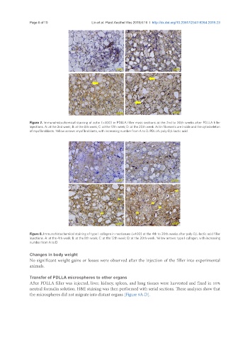
Figure 7. Immunohistochemical staining of actin (×400) in PDLLA filler mass sections at the 2nd to 20th weeks after PDLLA filler
injections. A: at the 2nd week; B: at the 8th week; C: at the 12th week; D: at the 20th week. Actin filaments are inside and the cytoskeleton
of myofibroblasts. Yellow arrows: myofibroblasts, with increasing number from A to D. PDLLA: poly-D,L-lactic acid
A B
C D
Figure 8. Immunohistochemical staining of type I collagen in neotissues (×400) at the 4th to 20th weeks after poly-D,L-lactic acid filler
injections. A: at the 4th week; B: at the 8th week; C: at the 12th week; D: at the 20th week. Yellow arrows: type I collagen, with increasing
number from A to D
Changes in body weight
No significant weight gains or losses were observed after the injection of the filler into experimental
animals.
Transfer of PDLLA microspheres to other organs
After PDLLA filler was injected, liver, kidney, spleen, and lung tissues were harvested and fixed in 10%
neutral formalin solution. H&E staining was then performed with serial sections. These analyses show that
the microspheres did not migrate into distant organs [Figure 9A-D].