
Page 105 - Read Online
P. 105
Page 6 of 9 Antunes et al. Plast Aesthet Res 2018;5:11 I http://dx.doi.org/10.20517/2347-9264.2018.03
Figure 9. Waters view radiography showing opacification of the right maxillary sinus
A B
C D
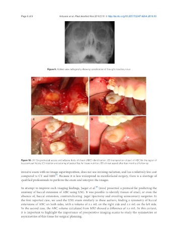
Figure 10. (A) Gingivobuccal access and adipose body of cheek (ABC) identification; (B) bransposition of part of ABC for the region of
buccosinusal fistula; (C) rotation and suturing of palatal flap for tissue nutrition; (D) clinical aspect after four months of follow-up
invasive exam with no image superimposition, does not use ionizing radiation, and has a relatively low cost
[8]
compared to CT and MRI . Because it is less widespread in maxillofacial surgery, there is a shortage of
qualified professionals to perform the exam and interpret the images.
[9]
In attempt to improve such imaging findings, Jaeger et al. (2016) presented a protocol for predicting the
anatomy of buccal extension of ABC using USG. It was possible to identify tissues of small, or even the
absence of, buccal extension, contraindicating jugal lipectomy and avoiding unnecessary surgeries. In
the first reported case, we used the USG exam similarly to these authors, finding a symmetry of buccal
extensions of ABC on both sides, with a volume of 2.4 mL on the right side and 2.2 mL on the left side.
In the second case, the ABC volume calculated from MRI showed a difference of 1.5 mL. In this context,
it is important to highlight the importance of preoperative imaging exams to study the symmetries or
asymmetries of this tissue for surgical planning.