
Page 101 - Read Online
P. 101
Page 2 of 9 Antunes et al. Plast Aesthet Res 2018;5:11 I http://dx.doi.org/10.20517/2347-9264.2018.03
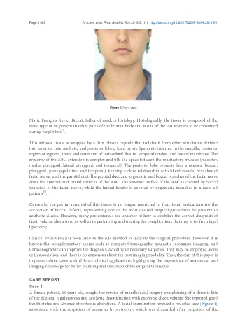
Figure 1. Front view
Marie François Xavier Bichat, father of modern histology. Histologically, the tissue is composed of the
same type of fat present in other parts of the human body and is one of the last reserves to be consumed
[2]
during weight loss .
This adipose tissue is wrapped by a thin fibrous capsule that isolates it from other structures, divided
into anterior, intermediate, and posterior lobes, fixed by six ligaments inserted in the maxilla, posterior
region of zygoma, inner and outer rim of infraorbital fissure, temporal tendon, and buccal membrane. The
anatomy of the ABC extension is complex and fills the space between the masticatory muscles (masseter,
medial pterygoid, lateral pterygoid, and temporal). The posterior lobe presents four processes (buccal,
pterygoid, pterygopalatine, and temporal), keeping a close relationship with blood vessels, branches of
facial nerve, and the parotid duct The parotid duct and zygomatic and buccal branches of the facial nerve
cross the anterior and lateral surfaces of the ABC. The anterior surface of the ABC is covered by buccal
branches of the facial nerve, while the lateral border is covered by zygomatic branches in almost all
[3]
patients .
Currently, the partial removal of this tissue is no longer restricted to functional indications for the
correction of buccal defects, representing one of the most desired surgical procedures by patients in
aesthetic clinics. However, many professionals are unaware of how to establish the correct diagnosis of
facial volume alterations, as well as in performing and treating the complications that may arise from jugal
lipectomy.
Clinical evaluation has been used as the sole method to indicate the surgical procedure. However, it is
known that complementary exams such as computed tomography, magnetic resonance imaging, and
ultrasonography can improve the diagnosis, avoiding unnecessary surgeries. They may be employed alone
or in association, and there is no consensus about the best imaging modality. Thus, the aim of this paper is
to present three cases with different clinical applications, highlighting the importance of anatomical and
imaging knowledge for better planning and execution of the surgical technique.
CASE REPORT
Case 1
A female patient, 24 years old, sought the service of maxillofacial surgery complaining of a chronic bite
of the bilateral jugal mucosa and aesthetic dissatisfaction with excessive cheek volume. She reported good
health status and absence of systemic alterations. A facial examination revealed a rounded face [Figure 1]
associated with the suspicion of masseter hypertrophy, which was discarded after palpation of the