
Page 104 - Read Online
P. 104
Antunes et al. Plast Aesthet Res 2018;5:11 I http://dx.doi.org/10.20517/2347-9264.2018.03 Page 5 of 9
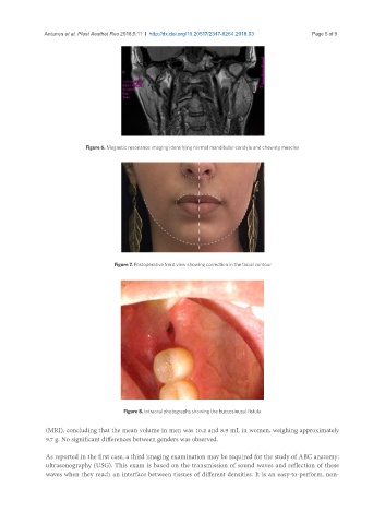
Figure 6. Magnetic resonance imaging identifying normal mandibular condyle and chewing muscles
Figure 7. Postoperative front view showing correction in the facial contour
Figure 8. Intraoral photographs showing the buccosinusal fistula
(MRI), concluding that the mean volume in men was 10.2 and 8.9 mL in women, weighing approximately
9.7 g. No significant differences between genders was observed.
As reported in the first case, a third imaging examination may be required for the study of ABC anatomy:
ultrasonography (USG). This exam is based on the transmission of sound waves and reflection of these
waves when they reach an interface between tissues of different densities. It is an easy-to-perform, non-