
Page 103 - Read Online
P. 103
Page 4 of 9 Antunes et al. Plast Aesthet Res 2018;5:11 I http://dx.doi.org/10.20517/2347-9264.2018.03
A B
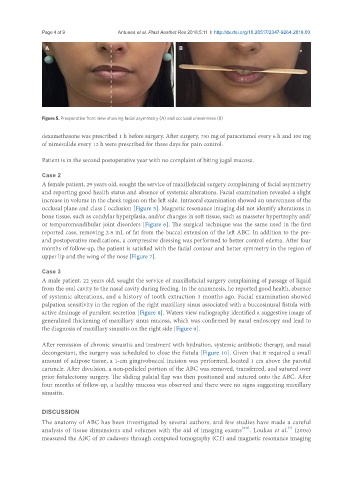
Figure 5. Preoperative front view showing facial asymmetry (A) and occlusal unevenness (B)
dexamethasone was prescribed 1 h before surgery. After surgery, 750 mg of paracetamol every 6 h and 100 mg
of nimesulide every 12 h were prescribed for three days for pain control.
Patient is in the second postoperative year with no complaint of biting jugal mucosa.
Case 2
A female patient, 29 years old, sought the service of maxillofacial surgery complaining of facial asymmetry
and reporting good health status and absence of systemic alterations. Facial examination revealed a slight
increase in volume in the cheek region on the left side. Intraoral examination showed an unevenness of the
occlusal plane and class I occlusion [Figure 5]. Magnetic resonance imaging did not identify alterations in
bone tissue, such as condylar hyperplasia, and/or changes in soft tissue, such as masseter hypertrophy and/
or temporomandibular joint disorders [Figure 6]. The surgical technique was the same used in the first
reported case, removing 2.8 mL of fat from the buccal extension of the left ABC. In addition to the pre-
and postoperative medications, a compressive dressing was performed to better control edema. After four
months of follow-up, the patient is satisfied with the facial contour and better symmetry in the region of
upper lip and the wing of the nose [Figure 7].
Case 3
A male patient, 22 years old, sought the service of maxillofacial surgery complaining of passage of liquid
from the oral cavity to the nasal cavity during feeding. In the anamnesis, he reported good health, absence
of systemic alterations, and a history of tooth extraction 3 months ago. Facial examination showed
palpation sensitivity in the region of the right maxillary sinus associated with a buccosinusal fistula with
active drainage of purulent secretion [Figure 8]. Waters view radiography identified a suggestive image of
generalized thickening of maxillary sinus mucosa, which was confirmed by nasal endoscopy and lead to
the diagnosis of maxillary sinusitis on the right side [Figure 9].
After remission of chronic sinusitis and treatment with hydration, systemic antibiotic therapy, and nasal
decongestant, the surgery was scheduled to close the fistula [Figure 10]. Given that it required a small
amount of adipose tissue, a 1-cm gingivobuccal incision was performed, located 1 cm above the parotid
caruncle. After divulsion, a non-pedicled portion of the ABC was removed, transferred, and sutured over
prior fistulectomy surgery. The sliding palatal flap was then positioned and sutured onto the ABC. After
four months of follow-up, a healthy mucosa was observed and there were no signs suggesting maxillary
sinusitis.
DISCUSSION
The anatomy of ABC has been investigated by several authors, and few studies have made a careful
[7]
analysis of tissue dimensions and volumes with the aid of imaging exams [4-6] . Loukas et al. (2006)
measured the ABC of 20 cadavers through computed tomography (CT) and magnetic resonance imaging