
Page 102 - Read Online
P. 102
Antunes et al. Plast Aesthet Res 2018;5:11 I http://dx.doi.org/10.20517/2347-9264.2018.03 Page 3 of 9
A B
Figure 2. Intraoral photographs on the right (A) and left (B) sides showing keratinization lines
A B
Figure 3. Doppler ultrasonography on the right (A) and left (B) sides, showing blood vessels over the surgical area
A B
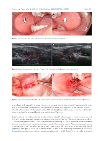
Figure 4. Intrabuccal photographs from the right side showing the adipose body of cheek identification (A) and link of blood vessels (B)
musculature and request for imaging exams. An intrabuccal examination revealed the presence of a white
line of keratinization, asymptomatic, parallel to the occlusion line, suggestive of an alba line [Figure 2].
Doppler ultrasound revealed symmetry of the ABC on the right and left sides and a very close relationship
of the branches of arteries and buccal veins on both sides [Figure 3].
Jugal lipectomy was performed under local anesthesia, using 2% lidocaine and 1:100,000 epinephrine, and
a parallel incision was made immediately adjacent to the mucosal bite line. The access distance was at least
1 cm below the parotid caruncle and could have been extended up to 2 cm. It is recommended to start the
incision 1 cm behind the caruncle, avoiding trauma in the masseter muscle. After submucous divulsion,
a portion of the ABC was identified along with blood vessels, which were linked with 4-0 silk thread
[Figure 4]. Next, part of the buccal extension of the ABC was pulled by rotating movements by a Halstead
forceps to remove the tissue and the mucosa was sutured with 3-0 silk thread. To prevent edema, 8 mg of