
Page 17 - Read Online
P. 17
Hwang et al. In vivo degradation of PLDLA-TMC
INTRODUCTION reduced. The floor and medial wall were reconstructed
with a trimmed (45 mm × 33 mm × 1.5 mm) CPS sheet
Absorbable sheets are used to treat orbital fractures. made of PLDLA-TMC (Inion Co., Tampere, Finland)
CPS sheets [poly L-lactic D-lactic acid and trimethylene [Figure 2].
carbonate (PLDLA-TMC)] absorbable sheets (Inion Co.,
Tampere, Finland) are commonly used to treat trauma Exophthalmometry on postoperative day (POD) 9
and in reconstructive procedures of the orbital cavity. revealed a 3-mm difference (right eye, 19 mm; left eye,
In vivo degradation of several absorbable materials 16 mm). The differences in exophthalmometry on POD
has been documented. [1-4] However, the compositional 98 and POD 154 were 7 mm (right eye, 18 mm; left eye,
changes (appearance of radicals, etc.) of PLDLA-TMC 11 mm) and 8 mm (right eye, 19 mm; left eye, 11 mm),
after insertion into the human body have not yet been respectively. On POD 189 (1 day before the secondary
described. operation), the difference was 5 mm (right eye, 18 mm;
left eye, 13 mm).
The aim of this paper was to assess compositional
changes in a PLDLA-TMC sheet, synthesized by a ring- Secondary reconstruction of the orbital floor was
opening polymerization process, 190 days after insertion performed with an iliac bone graft 190 days after
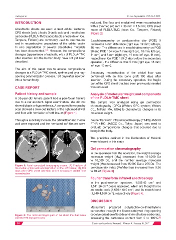
into a human body. insertion. During the secondary operation, the fragile
part of the CPS sheet that had been previously inserted
CASE REPORT was removed.
Patient history and sample Analysis of molecular weight and components
A 35-year-old female patient had a pan-facial fracture of the PLDLA-TMC sheet
due to a car accident. Upon examination, she did not The sample was analyzed using gel permeation
show diplopia or hypoesthesia. A computed tomography chromatography (GPC) (Waters GPC system; Waters
scan showed a blow-out fracture of the right medial wall Co., Milford, MA, USA) to characterize changes in its
and floor with herniation of soft tissues [Figure 1]. molecular weight.
Through a subciliary incision, the orbital floor and medial Fourier transform infrared spectroscopy (FT-IR) (JASCO
wall were exposed and the herniated soft tissues were FT-IR 4100; JASCO Co., Tokyo, Japan) was used to
evaluate compositional changes that occurred due to
A B being in the body.
The principles outlined in the Declaration of Helsinki
were followed in this study.
Gel permeation chromatography
In the specimen from the operation, the weight average
molecular weight (Mw) decreased from 151,000 Da
to 10,000 Da, and the number average molecular
weight (Mn) decreased from 15,600 Da to 255 Da. The
Figure 1: Axial computed tomography scans. (A) Fracture of
the medial orbital wall and herniation of the soft tissue; (B) 190 polydispersity index (Mn/Mn) thus increased from 9.96
days after CPS sheet insertion before secondary orbital floor to 40.22 [Figure 3].
reconstruction
Fourier transform infrared spectroscopy
In the post-insertion spectrum, 1,655.43 cm and
-1
1,541.26 cm peaks appeared, which are thought to be
-1
an amide peak (1,670-1,640 cm ) and its stretch bend
-1
(1,640-1,550 cm ), respectively [Figure 4].
-1
DISCUSSION
Matsumura prepared poly(lactide-co-trimethylene
carbonate) through the lipase-catalyzed ring-opening
Figure 2: The removed fragile part of the sheet that had been copolymerization of lactide and trimethylene carbonate,
inserted 190 days previously increasing the carbonate content from 0 to 100%. [5]
10 Plastic and Aesthetic Research ¦ Volume 4 ¦ January 19, 2017