
Page 139 - Read Online
P. 139
Ruiz-Moya et al. Assessment of DIEP flap using CTA with 3D reconstruction
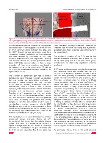
Figure 6: Three-dimensional abdominal wall reconstruction with AYRA software from computed tomography angiography images. (A)
Point of measurement (circle) of flap subcutaneous tissue thickness, at the level of the iliac crests horizontally and at the midpoint of rectus
abdominis width vertically; (B) measurement of flap subcutaneous tissue thickness
outflow from the superficial towards the deep system, show superficial drainage dominance. However, no
several studies [6,7,11] have suggested that the absence evidence was reported supporting this hypothesis.
of direct communications between the SIEV and Statistically significant evidence was neither found in
the DIEV through venous perforators could favor the present study.
congestion. However, statistically significant evidence
was only reported by Schaverien et al. using The number of branches of the SIEV was the last
[7]
magnetic angioresonance, and only when a DIEP flap anatomical feature evaluated, with a mean of 1.43
was dissected based on just one perforator without for the case group and 1.24 for the control group.
direct SIEV-DIEV communication. In fact, a larger Unfortunately, no statistically significant evidence
proportion of these communications was found in was found.
the case group than in the control group (57.14% vs.
38.10%) in the present study, although statistically not DIEP breast autologous reconstruction is recognized
significant (P = 0.42). as a reliable procedure with excellent outcomes and
low donor site morbidity. Whereas success rates of
[3]
The number of perforators per flap is another over 95% have generally been reported, some flaps
controversial topic. Previous research has revealed exhibit vascular complications and eventually fail.
[3]
that one medial row periumbilical perforator of The major complication that may arise is the diffuse
appropriate caliber provides the best perfusion venous congestion due to drainage insufficiency, [6,7]
to the DIEP flap including Hartrampf’s zone IV. [4,5] neither originated by venous thrombosis nor by
Nevertheless, from the point of view of venous technical failures. Finding predictive factors of
perfusion, DIEP flaps sometimes exhibit a diminished congestion preoperatively would be extremely helpful
drainage with an increased venous pressure. for the surgeon. Using duplex ultrasonography,
Douglas et al. suggested that just one arterial Figus et al. reported that the identification of the
[5]
[20]
perforator could provide the optimal perfusion, stating dominant venous perforator of the flap entailed
that with two arterial perforators the filling pressure high possibilities of finding an arterial perforator of
could drop, decreasing the gradient and favoring adequate caliber (93.5%), higher than the possibilities
congestion. For their part, Mohan et al. found a of finding a venous perforator of good caliber after
[18]
non-significant four-fold congestion rate in DIEP the identification of the dominant arterial perforator
flaps based on a single perforator compared to those (69.8%). Gravvanis et al. compared two subgroups
[21]
based on multiple perforators. In the present study, of breast reconstructions regarding vascular
no statistically significant evidence was found for the dissection: dominant arterial perforator-dissected
number of perforators per flap (P = 0.25). versus dominant venous perforator-dissected DIEPs. A
significant higher rate of venous congestion was found
The flap subcutaneous tissue thickness was another in the arterial perforator group. Laporta et al. and
[22]
anatomical feature analyzed. Rubino et al. Santanelli et al. [23] selected the type of perforators and
[19]
demonstrated that bigger flaps intrinsically develop their number for each flap depending on the diameter
greater flow rates, and consequently, demand a of the vein, and found that medial row perforators
higher drainage. Bast et al. found a correlation were a negative predictor for flap complications.
[14]
between the suprascarpal fat pad thickness and
the SIEV caliber, suggesting that thicker pads may As previously stated, CTA is the gold standard
132 Plastic and Aesthetic Research ¦ Volume 4 ¦ August 21, 2017