
Page 137 - Read Online
P. 137
Ruiz-Moya et al. Assessment of DIEP flap using CTA with 3D reconstruction
at the level of the most superior aspect of the iliac of both SIEVs across the abdominal midline were
crests and at the midpoint of the rectus abdominis found in 42.86% of flaps (3 cases), with a mean
muscle width [Figure 6]. diameter of the SIEV of 3.04 mm (± 0.60 mm), a
mean of 1.43 branches per SIEV, a mean of 1.86 (±
Statistical analysis 0.69) perforators nourishing each flap, and with an
According to the small sample size, the quantitative average flap subcutaneous tissue thickness of 3.56
variables were evaluated with the U-Mann-Whitney cm (± 0.90 cm) [Table 1]. In every congestive flap, an
non-parametric test, and the qualitative variables additional venous anastomosis was performed, either
with the Fisher exact test. For the statistical analysis, to the second concomitant vein of the DIEA (5 cases)
the IBM SPSS Statistics 19 package (SPSS Inc. or to the cephalic vein (2 cases). After this salvage
®
Chicago, IL) was used, considering significant procedure, all of the 7 flaps overcame congestion and
differences when P < 0.05. survived without necrosis. In the control group, direct
communications between the DIEA and the SIEV
RESULTS through perforators were found in 38.10% of flaps (8
controls), direct communications of both SIEVs across
The global venous congestion rate was 4.14% (7 the abdominal midline were found in 23.81% of flaps
flaps). The mean age of case and control subjects was (5 controls), with a mean diameter of the SIEV of 3.08
50.1 years (range 38-58 years) and 49.1 years (range mm (± 1.20 mm), a mean of 1.24 branches per SIEV, a
35-64 years), respectively. mean of 2.24 (± 0.77) perforators nourishing each flap,
and with a mean flap subcutaneous tissue thickness of
In the case group, direct communications between the 3.72 cm (± 0.83 cm) [Table 2]. No statistically significant
DIEA and the SIEV through perforators were found differences were found between the two groups for any
in 57.14% of flaps (4 cases), direct communications of the variables (P > 0.05) [Table 3].
DISCUSSION
The present study was not able to confirm any of the
studied anatomical variables as predictive factors
of venous congestion, despite being suggested
in the literature. [5,6,13,14] The abdominal superficial
venous dominance is one of the most extended and
accepted (but not proved) hypothesis for explaining
the diffuse congestion as a large diameter SIEV may
denote dominance over the deep venous system.
[6]
Blondeel et al. suggested that when this diameter
[13]
is > 1.5 mm, the SIEV should be preserved for venous
supercharging in case of congestion. However, in a
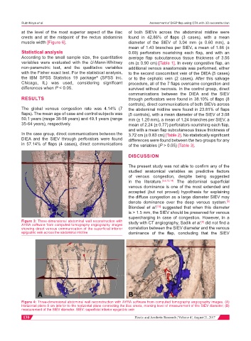
Figure 3: Three-dimensional abdominal wall reconstruction with study with CT angiography, Sadik et al. did not find a
[8]
AYRA software from computed tomography angiography images
showing direct venous communication of the superficial inferior correlation between the SIEV diameter and the venous
epigastric vein across the abdominal midline dominance of the flap, concluding that the SIEV
Figure 4: Three-dimensional abdominal wall reconstruction with AYRA software from computed tomography angiography images. (A)
Horizontal plane 8 cm inferior to the horizontal plane connecting the iliac crests, marking level of measurement of the SIEV diameter; (B)
measurement of the SIEV diameter. SIEV: superficial inferior epigastric vein
130 Plastic and Aesthetic Research ¦ Volume 4 ¦ August 21, 2017