
Page 138 - Read Online
P. 138
Ruiz-Moya et al. Assessment of DIEP flap using CTA with 3D reconstruction
Table 1: Variables studied in case group
Case Communication Communication Diameter of Branches of Perforators Subcutaneous Age
No. SIEV DIEA SIEV SIEV (mm) SIEV thickness (cm) (years)
1 No No 2.7 2 3 2.38 51
2 Yes Yes 3.6 2 2 3.19 48
3 Yes Yes 2.4 1 2 2.91 49
4 No No 2.9 1 2 4.49 38
5 Yes No 3.9 1 2 2.98 54
6 Yes Yes 3.5 2 1 4.35 53
7 No No 2.3 1 1 4.64 58
SIEV: superficial inferior epigastric vein; DIEA: deep inferior epigastric artery
Table 2: Variables studied in control group
Case Communication Communication Diameter of Branches of Perforators Subcutaneous Age
No. SIEV DIEA SIEV SIEV (mm) SIEV thickness (cm) (years)
1 Yes No 3.4 1 3 3.98 58
2 No No 3.0 1 3 2.72 54
3 No No 3.7 2 2 5.00 39
4 Yes No 2.7 1 2 4.14 57
5 No No 4.5 1 3 4.66 41
6 No No 1.7 1 2 3.70 64
7 Yes No 2.6 1 3 2.69 51
8 No No 3.1 1 2 4.50 35
9 No No 2.1 1 3 3.27 40
10 No Yes 2.4 2 2 3.50 50
11 Yes Yes 3.5 1 2 3.70 51
12 Yes No 3.6 2 2 4.42 47
13 No No 3.1 1 2 3.70 57
14 Yes No 3.3 2 1 4.27 50
15 No No 2.7 1 4 2.20 60
16 Yes Yes 3.9 1 2 3.57 37
17 No No 5.2 1 2 3.40 52
18 No No 2.1 1 1 5.49 36
19 Yes Yes 6.2 1 2 2.99 59
20 No Yes 3.0 2 1 3.57 50
21 No No 2.5 1 3 2.64 44
SIEV: superficial inferior epigastric vein; DIEA: deep inferior epigastric artery
Table 3: Statistical analysis of variables between groups
Cases Controls Significance
Variables Difference and 95% CI
(n = 7) (n = 21) (P)
Diameter of SIEV (mm), mean ± SE 3.04 ± 0.63 3.08 ± 1.22 0.915 -0.04 (-1.04, 0.95)
Branches of SIEV (2 branches), n (%) 3 (42.86) 5 (23.81) 0.371 19.05 (21.90, 60.00)
Perforators per flap, mean ± SE 1.86 ± 0.69 2.24 ± 0.77 0.255 -0.38 (-1.05, 0.29)
Subcutaneous thickness (cm), mean ± SE 3.56 ± 0.90 3.72 ± 0.83 0.652 -0.16 (-0.92, -0.60)
Communication SIEV-perforators, n (%) 4 (57.14) 8 (38.10) 0.418 19.05 (-23.10, 61.20)
Communication SIEVs midline, n (%) 3 (42.86) 5 (23.81) 0.371 19.05 (-21.90, 60.00)
SIEV: superficial inferior epigastric vein; CI: confidence interval; SE: standard error
diameter was not useful for predicting congestion.
This finding is consistent with the present study, as no
statistically significant evidence (P = 0.91) was found
when evaluating the SIEV diameter.
Another proposed feature in studies by Schaverien et al.,
[4]
Rozen et al., and Blondeel et al. was the absence
[6]
[13]
of direct venous communications of both SIEVs across
the abdominal midline, that could favor congestion
further this line. This hypothesis was not consistent with
the results of our study, as no statistically significant
evidence (P = 0.37) was found for this variable, being
these communications more numerous in the case
group than in the control group (48.86% vs. 23.81%).
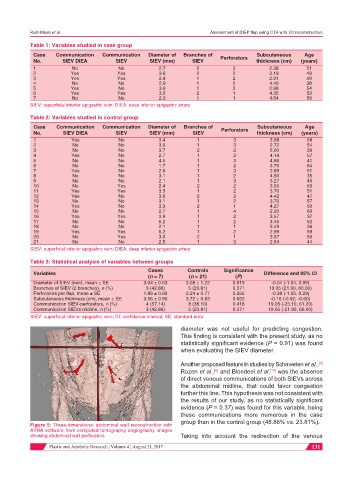
Figure 5: Three-dimensional abdominal wall reconstruction with
AYRA software from computed tomography angiography images
showing abdominal wall perforators Taking into account the redirection of the venous
Plastic and Aesthetic Research ¦ Volume 4 ¦ August 21, 2017 131