
Page 367 - Read Online
P. 367
Wang et al. Retroauricular skin/fascia expansion for microtia reconstruction
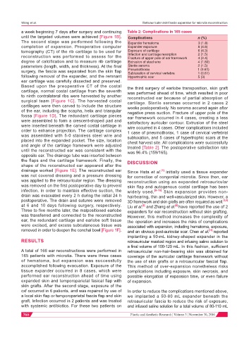
a week beginning 7 days after surgery and continuing Table 2: Complications in 165 cases
until the targeted volumes were achieved [Figure 1B]. Complications n (%)
The second stage was performed following the Expander hematoma 3 (1.8)
completion of expansion. Preoperative computer Expander exposure 8 (4.8)
tomography (CT) of the rib cartilage to be used for Exposure of cartilage 6 (4.3)
reconstruction was performed to assess for the Infection and cartilage resorption 2 (1.5)
Fracture of upper pole of ear framework 4 (2.4)
degree of calcification and to measure rib cartilage Extrusion of steel wire 4 (1.80)
parameters (length, width, and thickness). At the final Sterile seroma 2 (1.5)
surgery, the fascia was separated from the skin flap Pneumothorax 1 (0.61)
Subluxation of cervical vertebra 1 (0.61)
following removal of the expander, and the remnant Hypertrophic scar 5 (3)
ear cartilage was carefully dissected and preserved.
Based upon the preoperative CT of the costal the third surgery of earlobe transposition, skin graft
cartilage, normal costal cartilage from the seventh was performed ahead of time, which resulted in poor
to ninth contralateral ribs were harvested by another reconstructed ear because of partial absorption of
surgical team [Figure 1C]. The harvested costal cartilage. Sterile seromas occurred in 2 cases 2
cartilages were then carved to include the structure weeks postoperatively. No seroma accured again after
of the ear, including the scapha, helix and triangular negative press suction. Fracture of upper pole of the
fossa [Figure 1D]. The redundant cartilage pieces ear framework occurred in 4 cases, creating a less
were assembled to form a crescent-shaped pad and satisfactory auricular contour. Extrusion of the steel
were inserted beneath the carved costal cartilage in wire occurred in 4 cases. Other complications included
order to enhance projection. The cartilage complex 1 case of pneumothorax, 1 case of cervical vertebral
was assembled with 5-0 stainless steel wire and subluxation, and 5 cases of hypertrophic scars at the
placed into the expanded pocket. The size, location chest harvest site. All complications were successfully
and angle of the cartilage framework were adjusted treated [Table 2]. The postoperative satisfaction rate
until the reconstructed ear was consistent with the was 96.4% (159/165).
opposite ear. The drainage tube was inserted between
the flaps and the cartilage framework. Finally, the DISCUSSION
shape of the reconstructed ear appeared after the
drainage worked [Figure 1E]. The reconstructed ear Since Hata et al. initially used a tissue expander
[7]
was not covered dressing and a pressure dressing for correction of congenital microtia. Since then, ear
was applied to the retroauricular region. The dressing reconstruction using an expanded retroauricular
was removed on the first postoperative day to prevent skin flap and autogenous costal cartilage has been
infection. In order to maintain effective suction, the widely used. [8-10] Skin expansion provides non-
drain was evacuated every 2 h during the initial 24 h hairbearing, thin and well-vascularized skin. However, a
postoperative. The drain and sutures were removed 3D framework and skin grafts are often required as well. [8,9]
at 6 and 10 days following surgery, respectively. Liu et al. and Zhang et al. have reported the use of 2
[5]
[6]
Three to five months later, the malpositioned earlobe expanders for ear reconstruction without skin grafting.
was transferred and connected to the reconstructed However, this method increases the complexity of
ear, the redundant cartilage and earlobe soft tissue the operation and increases the risks of complications
were excised, and excess subcutaneous tissue was associated with expansion, including hematoma, exposure,
removed in order to deepen the conchal bowl [Figure 1F]. and an obvious post-auricular scar. Chen et al. [11] reported
implanting a 50-mL kidney-shaped expander in the
RESULTS retroauricular mastoid region and infusing saline solution to
a final volume of 100-120 mL. In this fashion, sufficient
A total of 166 ear reconstructions were performed in retroauricular non-hair-bearing skin was obtained for
165 patients with microtia. There were three cases coverage of the auricular cartilage framework without
of hematoma, but expansion was successfully the use of skin grafts or a retroauricular fascial flap.
accomplished following evacuation. Exposure of the This method of over-expansion nonetheless risks
tissue expander occurred in 8 cases, which were complications including exposure, skin necrosis, and
performed ear reconstruction ahead of time using possible elongation of expansion time, or even failure
expanded skin and temporoparietal fasical flap with of expansion.
skin grafts. After the second stage, exposure of the
cxf occurred in 6 patients, and was repaired by use of In order to reduce the complications mentioned above,
a local skin flap or temporoparietal fascia flap and skin we implanted a 50-80 mL expander beneath the
graft. Infection occurred in 2 patients and was treated retroauricular fascia to reduce the risk of exposure,
with systemic antibiotics. For these two patients on and infused saline solution for a total volume of 80-110 mL
366 Plastic and Aesthetic Research ¦ Volume 3 ¦ November 30, 2016