
Page 366 - Read Online
P. 366
Wang et al. Retroauricular skin/fascia expansion for microtia reconstruction
when a large series of ear reconstructions with tissue Table 1: Basic information of 165 patients (166 ears)
expansion were reported. [5,6] At present, retroauricular Characteristics n (%)
skin tissue expansion is one of the most popular Gender
techniques for ear reconstruction. In this study, tissue Male 112 (67.9)
expansion and autogenous costal cartilage were Female 53 (32.1)
Side
used for reconstruction of 165 patients with 166 Right 110 (66.7)
cases of microtia. The majority of the reconstructed Left 54 (33.3)
ears obtained a good contour with a low incidence of Bilateral 2 (0.60)
complications. In this paper, the authors report their Associated deformities 5 (3.6)
Hemifacial microsomia
experience with microtia reconstruction by use of full- Facial nerve weakness 1 (0.6)
thickness skin and fascia expansion and a three- Opposite ear deformity 2 (1.2)
dimensional costal cartilage framework. Fistula 4 (2.4)
3 (1.8)
Accessory ear
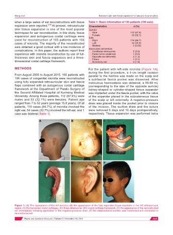
METHODS For the patient with left-side microtia [Figure 1A],
during the first procedure, a 4-cm length incision
From August 2005 to August 2015, 165 patients with parallel to the hairline was made on the scalp and
166 cases of congenital microtia were reconstructed a subfascial fascia pocket was dissected. After
using fully expanded retroauricular skin and fascia meticulous haemostasis was obtained, a 50-80 mL
flaps combined with an autogenous costal cartilage (corresponding to the size of the opposite auricle)
framework at the Department of Plastic Surgery of kidney-shaped or cylinder-shaped tissue expander
the Second Affiliated Hospital of Kunming Medical was implanted under the fascia pocket, with the valve
University. Among these patients, 112 (67.9%) were of the expander placed in the subcutaneous tissue
males and 53 (32.1%) were females. Patient age of the scalp or left externally. A negative-pressure
ranged from 7 to 52 years (average 15.8 years). Of all drain was placed inside the pocket prior to closure
patients, 110 cases (66.7%) of microtia involved the of the incision. The suction drain and the suture
right ear, 54 cases (32.7%) involved the left ear, and 1 were removed 6 days and 10 days postoperatively,
case was bilateral [Table 1]. respectively. Tissue expansion was performed twice
A B C
D E F
Figure 1: (A) The appearance of the left microtia; (B) the appearance of the fully expanded tissue expander in the left retroauricular
region; (C) the harvested costal cartilages; (D) three-dimensional (3D) costal cartilage framework; (E) the appearance of the reconstructed
ear immediately following application of the negative-pressure drain; (F) the malpositioned earlobe was transferred and connected to
reconstructed ear
Plastic and Aesthetic Research ¦ Volume 3 ¦ November 30, 2016 365