
Page 350 - Read Online
P. 350
Sabhalok et al. Epidermoid and dermoid cyst removal
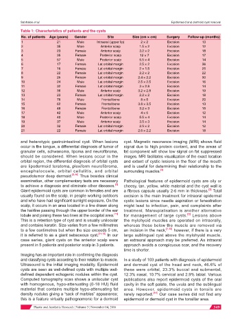
Table 1: Characteristics of patients and the cysts
No. of patients Age (years) Gender Site Size (cm × cm) Surgery Follow-up (months)
1 21 Male Intraoral (upper lip) 2 × 2 Excision 13
2 35 Male Anterior scalp 1.5 × 2 Excision 12
3 23 Female Anterior scalp 3.2 × 2 Excision 18
4 35 Female Posterior scalp 12 × 7 Excision 17
5 57 Male Posterior scalp 5.5 × 4 Excision 14
6 17 Female Lat orbital margin 2.5 × 2 Excision 36
7 16 Female Lat orbital margin 2 × 1.5 Excision 25
8 22 Female Lat orbital margin 2.2 × 2 Excision 22
9 25 Female Lat orbital margin 2.4 × 2.2 Excision 30
10 24 Male Lat orbital margin 2.5 × 2.5 Excision 16
11 32 Female Lat orbital margin 3 × 2.8 Excision 14
12 36 Male Anterior scalp 3.2 × 2.8 Excision 19
13 23 Female Lat orbital margin 2.2 × 2 Excision 18
14 78 Male Frontal bone 8 × 5 Excision 20
15 62 Female Frontal bone 3.8 × 3.5 Excision 13
16 48 Female Frontal bone 3.2 × 3 Excision 15
17 42 Male Anterior scalp 4 × 5 Excision 12
18 40 Male Posterior scalp 5.5 × 4 Excision 14
19 37 Male Anterior scalp 3.5 × 3 Excision 14
20 32 Female Lat orbital margin 2.5 × 2 Excision 12
21 22 Female Lat orbital margin 2.6 × 2.2 Excision 18
and heterotypic gastrointestinal cyst. When lesions cyst. Magnetic resonance imaging (MRI) shows fluid
occur in the tongue, a differential diagnosis of tumor of signal due to high protein content, and the areas of
granular cells, schwanoma, lipoma and neurofibroma, fat component will show low signal on fat suppressed
should be considered. When lesions occur in the images. MRI facilitates visualization of the exact location
orbital region, the differential diagnosis of orbital cysts and extent of cystic lesions in the floor of the mouth
are lipodermoid teratoma, plexiform neurofibroma, and is useful for determining their relationship to the
encephalocoele, orbital cellulitis, and orbital surrounding muscles. [3]
pseudotumor deep dermoid. [9,10] Thus besides clinical
examination, other complementary tests are necessary Pathological features of epidermoid cysts are oily or
[2]
to achieve a diagnosis and eliminate other diseases. cheesy, tan, yellow, white material and the cyst wall is
Giant epidermoid cysts are common in females and are a fibrous capsule usually 2-6 mm in thickness. Total
[3]
usually found on the scalp, in people working outdoors excision is the main treatment for intraoral epidermal
and who have had significant sunlight exposure. On the cystic lesions since needle aspiration or fenestration
scalp, it occurs in an area located in a line drawn along might lead to infection, pain, and complaints after
the hairline passing through the upper border of the ear treatment. Marsupialisation is another alternative
[11]
[3]
lobule and joining these two lines at the occipital area. for management of large cysts. Lesions above
This is a retention type of cyst and is usually unilocular the mylohyoid muscles are operated on intraorally,
and contains keratin. Size varies from a few millimetres whereas those below the muscle are removed via
to a few centimetres but when the size exceeds 5 cm, an incision in the neck, [1,13] however, if there is a very
it is referred to as a giant sebaceous cyst. [11,12] In our large sublingual cyst above the mylohyoid muscle,
case series, giant cysts on the anterior scalp were an extraoral approach may be preferred. An intraoral
present in 5 patients and posterior scalp in 3 patients. approach avoids a conspicuous scar, and the recovery
time is shorter.
Imaging has an important role in confirming the diagnosis
and classifying cysts according to their relation to muscle. In a study of 103 patients with diagnosis of epidermoid
Ultrasound is the initial imaging modality. Epidermoid and dermoid cyst of the head and neck, 46.6% of
cysts are seen as well-defined cysts with multiple well- these were orbital, 23.3% buccal and submental,
defined dependent echogenic nodules within the cyst. 12.3% nasal, 10.7% cervical and 2.9% labial. Various
Computed tomography scan shows a unilocular cyst publications also report epidermoid cysts of the oral
with homogenous, hypo-attenuating (0-18 HU) fluid cavity in the soft palate, the uvula and the sublingual
material that contains multiple hypo-attenuating fat area. However, epidermoid cysts in tonsils are
density nodules giving a “sack of marbles” appearance; rarely reported. [14] Our case series did not find any
this is a feature virtually pathognomonic for a dermoid epidermoid or dermoid cyst in the tonsillar area.
Plastic and Aesthetic Research ¦ Volume 3 ¦ November 04, 2016 349