
Page 349 - Read Online
P. 349
Sabhalok et al. Epidermoid and dermoid cyst removal
A B A B
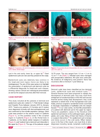
Figure 1: Preoperative (A) and intra-operative (B) view of a dermoid Figure 2: Preoperative (A) and intra-operative (B) view of a dermoid
cyst of scalp cyst of occiput
A B A B
Figure 3: Preoperative (A) and intra-operative (B) view of a dermoid Figure 4: Preoperative (A) and intra-operative (B) view of a epidermoid
cyst of lateral orbital margin cyst of the upper lip
[7]
cyst in the oral cavity, lower lip, or upper lip. Giant 16-78 years. The size ranged from 1.5 cm × 2 cm to
epidermoid cysts are rare and they present in the scalp. 12 cm × 7 cm [Table 1]. Infected cysts were managed
with preoperative antibiotics. All cysts were unilocular.
Epidermoid cysts are relatively less common in No evidence of malignancy was present. None had
the head and neck region, hence are likely to be recurrence after a minimum 1-year follow-up.
misdiagnosed. The aim of this case series is to highlight
the presentation of epidermoid and dermoid cysts as DISCUSSION
a differential diagnosis for head and neck masses, Dermoid cysts have been classified as true dermoid
showing various clinical and radiological presentations cysts, epidermoid cysts and teratoid cysts. [1-3,6]
as well as the surgical outcomes after their removal. Several theories have been proposed to explain the
development of dermoid cysts: they may result from
CASE REPORT entrapment of ectodermal tissue of the first and second
brachial arches during fetal development; they could
This study included all the patients of dermoid and represent a variant form of the thyroglossal duct cyst;
epidermoid cysts who visited D. Y. Patil Dental College finally, previous surgical or accidental events could lead
and Hospital, Pune between January 2010 to January to traumatic implantation of epithelial cells into deeper
2015. Twenty-one patients (12 females and 9 males) tissues. [1,4] In our case series, we had the presentation
were diagnosed clinically with dermoid/epidermoid cyst of a patient with dermoid cyst in the upper lip region.
and confirmed by fine-needle aspiration cytology. Giant There are paediatric cases in the literature with
cysts were present on the anterior scalp in 5 patients epidermal cyst lesions in the sublingual region, gingiva,
[Figure 1], on the posterior scalp in the occipital palate, and uvula. Many of them had history of trauma
region in 3 patients [Figure 2], on the frontal bone in or surgical intervention. An epidermoid cyst is benign
[8]
3 patients, on the lateral orbital margins in 9 patients and rarely occurs in the oral cavity. When lesions
[Figure 3], and 1patient had a dermoid cyst intraorally in occur in the floor of the mouth, one must think of other
the upper lip [Figure 4]. The age of the patients ranged diagnoses including ranula, lymphatic malformation
348 Plastic and Aesthetic Research ¦ Volume 3 ¦ November 04, 2016