
Page 342 - Read Online
P. 342
Haffner The temporal endoscopic midface lift
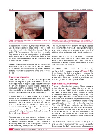
Figure 3: Mobilising of the midface by endoscopic dissection of Figure 4: Superficial musculoaponeurosis system suture and
the retaining ligaments imbrication under endoscopic control with the authors’ technique
enmeshed and reinforced by the fibres of the SMAS. The results are achieved primarily through the correct
Both the superficial and deep parts of the fat pad repositioning of the midface, the appropriate redraping
hold sutures well as the intertwined Camper’s fascia and vector rotation and anchorage of skin flap, all of
and SMAS fibres respectively, lend them strength. which are then well supported by SMAS imbrication.
This unique anatomical construct of the reinforced
malar fat pad serves as an effective pole for which Usage of an endoscope is mandatory. The author
lifting and anchoring threads can be secured to with advocates video recording of the procedure potentially
effectiveness and longevity. for accurate documentation in case review is
necessary in future. Precise haemostasis is better
The key elements of the method are the endoscopic with endoscopic visualization.
dissection in the superficial plane, the high malar
SMAS anchor, the direct internal flap anchor and the The SMAS must be imbricated to provide stability
author’s suture technique in the tunnel and keyhole similar to that of an open procedure [Figure 4]. This
access. is challenging due to the long distance between the
incision and imbrication sites. Furthermore, the poor
Endoscopic dissection ergonomics of the instrumentation requiring a key-
Once the plane of dissection has progressed hold like grip makes suturing more difficult.
beyond the zygoma, a rigid 4 mm diameter operative
endoscope with a 30-degree angle is used to visualise High malar SMAS anchorage
this area for dissection. The endoscopic dissector is The author advocates that the malar fat pad be viewed
introduced over the endoscope through the temporal not as a fat pad, which implies a flimsy structure, but
incision. A distal spoon-shaped shield is also used to instead as malar SMAS that has implies a stronger
maintain proper visualization of the optical cavity. structure. The justification for this nomenclature change
is due to the reinforced and sturdy nature of both parts
A hollow space is created using the endocopic of this fat pad described above. Furthermore, usage
dissector to progressively elevate the skin. The of the new term this way is key to understanding the
internal facial structures are visible throughout the role of the malar SMAS for midface repositioning,
dissection. The endpoint for a good working cavity special threads, special needles and anchoring points.
is reached after release of the fasciocutaneous The malar SMAS can be directly repositioned and
ligaments, especially the zygomatic and the parotido- anchored high on the zygomatic bone, approximately
masseteric ligaments. Attention must be paid to avoid 2 cm below the horizontal level of the corner of the eye
incorrectly identifying ligaments that are branches of in the middle third of the zygomatic bone to effect the
the facial nerves [Figure 3]. midface lift. This is what the author terms high malar
SMAS anchorage and is the cornerstone-working step
SMAS excision is not mandatory as good results can of the TEM.
already be achieved in conventional facelifts without
it. The most challenging element of the procedure is The author uses permanent sutures for this purpose
maintaining dissection in the correct plane. Adherence and specifically obtains informed patient consent for
to this avoids any damage to the facial nerve. this purpose. Braided, non-resorbable silicone-coated
Plastic and Aesthetic Research ¦ Volume 3 ¦ October 31, 2016 341