Page 337 - Read Online
P. 337

Sharma et al.                                                                                                                                                                                                 Morel-Lavalle lesion

            A                                                 A                       B


                                                                                             Flow
                                                                                             movements
            B


                                                              C


           Figure 1: Photograph of the right leg with focal swelling. (A) fronto-
           lateral position of the thigh shows swelling on upper lateral part with   No flow
           slight discoloration (white solid arrow); (B) magnified view of the same
           swelling shows normal overlying skin surface (hollow white arrow)
            A                      B
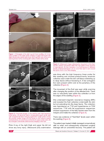
                                                              Figure 3: Ultrasound images showing the movement of the fluid.
                                                              (A) few uniform soft echoes within the fluid (white arrow) because
                                                              of haemolymph; (B) flow moments in the fluid because of different
                                                              density of blood and lymph; (C) no color flow seen in and around
                                                              the cycstic fluid in color flow imaging
                                                              was done with the high frequency linear probe for
                                                              this swelling and showed predominantly anechoic
                                                              collection seen under the skin soft tissue extending up
                                                              to deep fascia without traversing it. A few echogenic
                                                              foci were present within and at the periphery of the
                                                              collection [Figure 2].
            C                      D
                                                              The movement of the fluid was seen while scanning
                                                              after changing the position of the affected limb. There
                                                              was no vascularity seen within the collection or from
                                                              the edges of the swelling [Figure 3].

                                                              She underwent magnetic resonance imaging (MRI)
                                                              and revealed the fluid collection underneath the skin
                                                              but not extending into the deep fascia. The collection
                                                              was hypointense on T1W and hyperintense on T2W
                                                              sequences. T1W with fat suppression sequences
                                                              showed suppression of the fatty lobules seen from the
                                                              margins and inside the collection [Figure 4].
           Figure 2: Greyscale images of the swelling with high frequency
           linear probe. (A) anechoic fluid in encapsulated space (white star)
           with echogenic fat lobule hanging against the margin (white arrow);   There was evidence of “fluid-fluid” levels seen within
           (B) another similar image with fat lobule on the anterior aspect   the swelling [Figure 5].
           (upward arrow); (C) anechoic pure fluid in the space (white star); (D)
           two echogenic fat lobules (white arrows) hanging in the fluid
                                                              The patient was treated initially managed conservatively
           Plain X-ray of the right thigh and upper hip did not   with no relief, and subsequently underwent surgical
           show any bony injury. Ultrasound (US) examination   drainage with an uneventful recovery. The patient will

            336                                                                                       Plastic and Aesthetic Research ¦ Volume 3 ¦ October 25, 2016
   332   333   334   335   336   337   338   339   340   341   342