
Page 341 - Read Online
P. 341
Haffner The temporal endoscopic midface lift
longer recovery. The dissection plane has to be superficial layer of the temporal fascia. The dissecting
developed under the superficial temporal fascia and plane is then transitioned onto the superficial surface
then transitioned to the subperiosteal plane over the of the superficial layer of the temporal fascia.
zygoma. The surfaces of the zygoma and malar bone
then need to be connected through subperiosteal The subsequent surgical steps are common to both
dissection. Subsequent anchorage of the malar approaches. As dissection proceeds over the zygoma,
tissues are achieved only by suspension of the malar the temporal branch of the facial nerve remains
[8]
fat pad, and its longevity is questionable. Other protected, it is deep to the superficial layer of the
alternative procedures such as the minimal access temporal fascia. Both the mid-facial (malar) SMAS and
cranial suspension (MACS) lift with a third suture [10] the lateral facial SMAS can be easily reached this way.
cause a visible facial scar of 14-16 cm in length.
Pertinent anatomy
METHODS The temporal and facial portions of the SMAS fuse
over the malar bone. The facial SMAS continues in
The temporal endoscopic midface (TEM) lift is a new the temple as the superficial temporal fascia and
[1]
minimal-access facelift that uses exclusively temporal the frontal branch of the facial nerve lies deep to it.
access incisions thereby sparing any scars on the Limiting dissection to the plane above this fascia
face itself. This utilizes endoscopic dissection and a without violating it will ensure avoidance of nerve
[11]
suturing technique that was developed by the author. injury. The facial SMAS continues in the midface
[1]
An incision measuring 5-6 cm is made and hidden in as the malar SMAS and is incorporated by the thick
the hair-bearing part of the temple. Common pitfalls malar fat pad.
such as damaging the hair roots or making the flap
excessively thin must be avoided at this stage. The The anatomy of the facial fat compartments and that
dissection plane is developed over the superficial of the malar fat pad was best described by Botti and
surface of the common facial and temporal superficial Ceravolo through cadaveric studies. The malar
[1]
musculoaponeurotic system (SMAS). It is important to fat pad was found to be divided into two parts, a
completely avoid violating the integrity of the SMAS superficial part and a deeper part.
by either inadvertent incisions or diathermy.
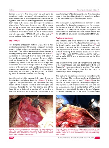
The author´s clinical experience is consistent with
An alternative initial approach through the same these findings. The midface can be well visualised
incision is a dual plane dissection [Figure 1]. In this through a medial extended facelift [Figure 2] and the
approach, the plane between the superficial and deep parts of the malar fat pad can be easily distinguished.
layers of the temporal fascia is first developed and The superficial part originates from the skin and can
dissected towards the non-hair bearing skin of the be conceptualised as a condensation of the malar
face. When or before the junction of the hairline is thickening of skin fat with the strong Camper´s fascia.
reached, an incision is made on the deep surface of the Under this, the deep part of the malar fat pad is found
mfp
SMAS
Figure 1: Dual plane temporal dissection. The superficial plane Figure 2: Demonstration of the midface with the malar fat pad
above the superficial fascia is exposed after dissecting the deep (malar SMAS) during an extended temporal-cervical-facial open
plane first. The superficial fascia is fixed by sutures onto the deeper facelift. SMAS: superficial musculoaponeurosis system; mfp:
one in order to facilitate the dissection malar fat pad
340 Plastic and Aesthetic Research ¦ Volume 3 ¦ October 31, 2016