
Page 40 - Read Online
P. 40
the current series. Although composite phalloplasty can of skin plasties can be planned in advance and performed
be performed under local anesthesia and sedation, the at the beginning of the procedure, the author prefers to do
author prefers spinal anesthesia that adds little morbidity this once the penis advancement has been completed, to
and enhances patient comfort. The procedure begins with modify for each situation. Treatment of the skin gap begins
a 3.5 cm incision located about 2.5-3.0 cm cranial to by closing the defect in a horizontal fashion [Figure 4]. This
the peneo‑pubic angle. The first step is to perform the closure produces two dog ears that will provide the final
dissection and release of the fascial and fasciocutaneous measure of skin advancement. The distal dog ear is tailored
attachments. The dissection then proceeds down to the to provide a Y or T advancement. The proximal dog ear is
front edge of the suspensory ligament. Thus, the release usually smaller and can be managed by defatting and direct
must be performed directly from the attachments to the closure; in about 2-3 months it will flat tenon on its own.
symphyseal ligament to avoid accidental damage to deep Performed correctly, closure of the skin by an advancement
penile neurovascular structures. The release is then carried plasty stabilizes and maintains the improvement in
further down, stopping at the start of the pelvic floor. The length [Figure 5]. It must be kept in mind that an overly
author usually does not release bone attachments except in ambitious cutaneous advancement usually results in
cases of micropenis. After the ligament release is complete, the incorporation of hairy skin and some scrotalization
corpora cavernosa will move easily forward and downward, of the penis shaft which worsens the aesthetic result.
creating a dead space between these structures and the Before epidermal closure, the author inserts a vacuum
pubic bone; This dead space must be filled with local drain and then proceeds to girth augmentation with fat
tissues; the availability of these tissues can be extremely grafting as previously described. All sutures used including
variable depending on the body mass index of the patient. epidermal closure can be performed with 4/0 absorbable
In slim patients it is usually necessary to take the fat that monofilament.
surrounds the spermatic cords. When there is enough As a rule composite augmentation phalloplasty can be
pubic fat, adipofascial flaps can be tailored and turned performed on an outpatient basis. The drain is removed
down as described by Hinderer and Espinosa. Available after 24 h and antibiotics are continued for 3 days. After
[4]
tissues are interposed inside the dead space created by the
ligament release while simultaneously pulling on the penis
and checking on the stability of the repair.
Upon completion of these steps, a skin gap can be observed
and that is caused by penis advancement. Although a variety
b
a
a b
c d
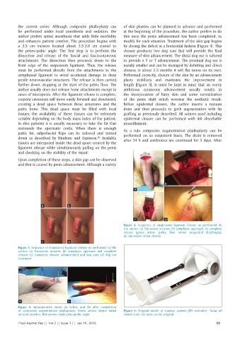
Figure 4: Sequence of suspensory ligament release as performed by
the author. (a) Transverse incision; (b) symphysis approach; (c) complete
release (green arrow: pubis, blue arrow: urogenital diaphragm);
(d) transverse initial closure
c d
Figure 3: Sequence of suspensory ligament release as performed by the
author. (a) Transverse incision; (b) symphysis approach and complete
release; (c) transverse closure, advancement and dog ears; (d) dog ear
treatment
a b
Figure 5: Intraoperative views (a) before and (b) after completion
of composite augmentation phalloplasty. Green arrows depict initial Figure 6: Original model of traction system (JES extender). Today all
incision location. Red arrows mark peneopubic angle brands look the same as the original
Plast Aesthet Res || Vol 2 || Issue 1 || Jan 15, 2015 29