
Page 322 - Read Online
P. 322
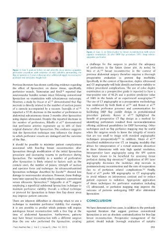
a                   b

                                                               a                        b
                                                              Figure  2: Case 2, (a) Patient prior to breast reconstruction with severe
                                                              capsular contracture;  (b) after  DIEP  flap procedure.  DIEP:  Deep  inferior
                                                              epigastric perforator


                                                              a challenge for the surgeon to predict the adequacy
                        c
                                                              of  perforators  in  the  future  donor  site.  As  noted  by
          Figure 1: Case 1, patient before (a) and after (b), deep inferior epigastric   Wes  et  al.,   breast reconstructive in the context of
                                                                        [21]
          perforator  procedure with evidence of mild cellulitis surrounding the
          flap; (c) patient at 2 years follow‑up after additional nipple reconstructive   previous  abdominal  surgery  therefore  requires  a  thorough
          produces and extensive weight loss                  preoperative  evaluation  to  prevent  flap  morbidity.
                                                              Specifically in the context of liposuction, duplex ultrasound
          Previous literature has shown conflicting evidence regarding   and CT angiography will help identify perforator viability to
          the  effect  of  liposuction  on  donor  tissue,  specifically,   reduce procedural complications. The use of color duplex
          perforator vessels. Teimourian and Kroll   reported that   examination as a preoperative guide is reported to have a
                                             [16]
          neurovascular bundles remain intact following conventional   true‑positive rate of 96.2% and a positive predictive value
          liposuction  on  examination  with  subcutaneous  endoscopy.   of 100% in the hands of an experienced sonographer.
                                                                                                             [22]
          However, a study by Ozcan et al.  demonstrated that flap   The use of CT angiography as a preoperative methodology
                                     [17]
          necrosis is directly related to the number of suction passes   was reinforced by both Bank  et  al.  and Rozen  et  al.
                                                                                            [23]
                                                                                                             [24]
                                                         [18]
          of a cannula accompanied by a vacuum. İnceoğlu et al.    to confirm perforator presence and communication for
          reported a 57.8% decrease in the number of perforators in   facilitating DIEP flap paddle design in postabdominal
                                                                                              [24]
          abdominal subcutaneous tissue 3 months after liposuction   procedure patients. Rozen  et  al.  highlighted the
          using duplex ultrasound. Despite the reported decrease in   benefit  of  preoperative  CT  flap  design  as  a  method  for
                                             [5]
          the number  of perforators, Ribuffo  et  al.  demonstrated   identifying perforators resulted from neovascularization
          that perforator arteries  regenerate up to 40%  of their   offering additional possibilities for DIEP harvesting. Other
          original diameter after liposuction. This evidence suggests   techniques such as flap perfusion mapping may be useful
          that the liposuction technique may influence the degree   when the surgeon needs to know the integrity of vessels
          to which perforator vessels are damaged and the outcome   that are too small to image with standard angiographic
                                                                                    [25]
                                                                                                             [26]
          of the flap.                                        techniques  preoperatively.   In  addition,  Masia  et  al.
                                                              described multidetector‑row CT, an imaging modality that
          It should be possible to minimize patient complications   allows for interpretation of a virtual anatomic dissection
          associated with free‑flap breast reconstruction after   in three dimensions with very high spatial resolution.
          liposuction through modification of the initial liposuction   Intraoperative laser angiography using the SPY system
          procedure and decreasing trauma to perforators during   has been shown to be beneficial for assessing tissue
          liposuction. The variability in a number of perforators   perfusion during flap elevation.  Application of SPY laser
                                                                                         [27]
          after  liposuction  is  likely related  to  factors  such  as the   angiography decreases the incidence skip necrosis in
          cannula  used,  the  number  of  passes,  strength  of  suction   postmastectomy reconstruction and the rate of reoperation
          and operator force and technique. An ultrasound‑assisted   due to of perfusion related complications.  Finally,
                                                                                                      [28]
          liposuction technique described by Zocchi  showed less   Farid  et  al.  prefer MR angiography to CT angiography
                                              [19]
                                                                        [8]
          damage to neurovascular structures. However, these findings   to avoid reliance on intravenous contrast and to reduce
          were later opposed by a study which compared conventional   patient exposure to radiation. Appropriate application
          versus ultrasonic liposuction.  Salgarello  et  al.  suggest   of  these  techniques  for  perforator  evaluation  including
                                   [2]
                                                  [20]
          employing a superficial subdermal liposuction technique to   CT, ultrasound, or perfusion mapping may improve the
          maintain perforator viability. Overall, a refined technique   outcome  of  patients  undergoing  DIEP after abdominal
          or protocol for liposuction in future free flap donor areas   liposuction
          may improve patient outcome.
          There are inherent difficulties in  choosing when to use a   CONCLUSION
          technique to maximize perforator viability. For example,
          it is not possible to predict which patients will require   We have demonstrated two cases, in addition to the previously
          autologous breast reconstruction with a free flap at the   reported literature that suggest previous conventional
          time of abdominal liposuction. Furthermore, patients   liposuction is not an absolute contraindication for free‑flap
          may have  breast reconstruction  with  a  different  surgeon   breast reconstruction. Preoperative management  of  the
          than the one who performed the liposuction, creating   patient  should  include  thorough  evaluation  of  suitable
          Plast Aesthet Res || Vol 2 || Issue 6 || Nov 12, 2015                                             313