
Page 325 - Read Online
P. 325
in certain surgical maneuvers. Patient safety is optimized to nasal tip should be aligned and straight. [14,15] Over
with the use of specific surgical procedures, protocols, reduction of the dorsum can change the orbito‑nasal
specialized instruments and staff training. Endotracheal relationship with subsequent flattening of the midface.
monitored anesthesia care is preferable and a
nasopharyngeal pack can be a useful preventative measure COMPLICATIONS OF RHINOPLASTY
by helping to keep the larynx clear.
Clinical manifestations of complications of rhinoplasty
THE AESTHETIC ANATOMY OF THE and side effects may be classified as functional,
NOSE: DORSAL AESTHETIC LINES aesthetic, or both. A number of technical solutions
have been presented. [4,5,12,16] After a review of these
The bony cartilaginous pyramid of the external nose is potential complications, specific attention was directed
three‑dimensional structure composed of three basic to the surgical technique for reconstruction of nasal tip
regions: the upper rigid bony third, the middle semi‑rigid projection and the dorsal aesthetic lines in the patient
cartilaginous third and the lower mobile cartilaginous with a prominent dorsal hump. Functional insufficiency
third. Nasal deformities result from the loss of support to of the internal nasal valve occurs in conjunction with
this tripod [Figure 1]. [4] the inverted V deformity (with disruption of the dorsal
aesthetic lines) caused by collapse of the upper lateral
The soft tissue components of the nose include skin, cartilages following removal of the dorsal hump.
muscles, nerves and vascular tissues. The tissue layers and This combined complication can be prevented during
fibrovascular membranous structures of the skin envelope component reduction of the dorsal hump by avoiding
in the inferior part of the external nose are divided into excessive resection of the upper lateral cartilage as
five layers, which are similar to the structure of the face: compared with the septum (midvault area) and by
epidermis, dermis, superficial fascia, fibromuscular layer placement of spreader grafts. [17]
and perichondrium. The thin, dynamic musculoaponeurotic
layer of the nose is a critical structure of the nose. The nasal tip presents an exceptional challenge because
Preservation of this layer is vital in restoring and retaining of its mobility. [12‑15] During dorsal hump reduction when
nasal function and appearance. [8‑10] the K‑area is disrupted and not aligned with the nasal
bridge, it may act as a pivot point; downward and inward
The nasal dorsum connects the radix to the lateral rotation of the septal cartilage then becomes possible,
projections of the crura of the lower lateral cartilages (LLCs) disproportionally widen the nasal dorsum and result
by means of two diverging concave lines. These are the unnatural look of dorsal aesthetic lines. Protrusion of
nasal dorsal aesthetic lines, which are unbroken extensions the anterior septal cartilage can create a polly beak
of the superciliary ridges [Figure 2]. The radix and supratip deformity. The polly beak deformity is remarkable for
regions have thicker soft tissue coverage, while the protuberance with a rounded downward pointing tip
midvault area contains thinner tissue. The supratip break and fullness of the supratip region. Excess scar tissue
occurs cephalad to the nasal tip where the contour lines in the region of the dorsal septal cartilage or supratip
of the nasal dorsum rise toward the tip‑defining points. may become apparent once edema has resolved and is
The tip‑defining points are composed of two equilateral more likely to occur in patients with thicker skin. The
triangles which extend from the supratip region to the deformity can be prevented by maintaining adequate tip
apex of the domes to the columellar lobule angle. [8,11,12] To support through columellar struts. In addition, suturing
achieve a balanced dorsal profile with a supratip break, the subcutaneous tissue of the supratip to the caudal
it is necessary to create a frame with a slightly deeper
nasion and tip projection beyond the dorsum. [10‑13] From
an aesthetic standpoint, the area from the nasal bridge
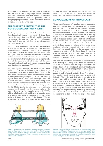
Figure 1: The keystone area, where the nasal bones overlap the upper Figure 2: The dorsal aesthetic lines originate on the supraorbital ridges
lateral cartilages and the scroll area, where the lower lateral cartilages and pass medially along the glabellar area to converge caudally at the
overlap the upper lateral cartilages. Restoration of the keystone area medial canthal ligaments. From there, they usually begin diverging at the
anatomical structure during the primary rhinoplasty prevents open roof keystone area and ultimately conclude at the tip‑defining points, which
and inverted V deformities become the highest point in the nasal profile
316 Plast Aesthet Res || Vol 2 || Issue 6 || Nov 12, 2015