
Page 274 - Read Online
P. 274
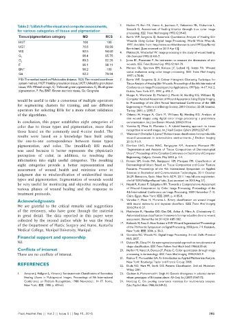
Table 2: %Match of the visual and computer assessments,   2.   Herbin M, Bon FX, Venot A, Jeanlouis F, Dubertret ML, Dubertret L,
          for various categories of tissue and pigmentation       Strauch  G.  Assessment  of  healing  kinetics  through  true  color  image
                                                                  processing. IEEE Trans Med Imaging 1993;12:39‑43.
           Tissue/pigmentation category    MD          RCS    3.   Berris WP, Sangwine SJ. Automatic Quantitative Analysis of Healing Skin
           HGT                             100         100        Wounds Using Colour Digital Image Processing. World Wide Wounds,
           UGT                             76.6        89.99      1997; Available from: http://www.worldwidewounds.com/1997/july/Berris/
                                                                  Berris.html. [Last accessed on 2015 Apr 12].
           WS                              80.5        94.60  4.   Mekkes JR, Westerhof W. Image processing in the study of wound healing.
           G 1                             98.4        95.75      Clin Dermatol 1995;13:401‑7.
           G 2                             89.3        92.26  5.   Jones BF, Plassmann P. An instrument to measure the dimensions of skin
           F                               96.1        98.11      wounds. IEEE Trans Biomed Eng 1995;42:464‑70.
           BNT                             98.2        100    6.   Hansen  GL,  Sparrow  EM,  Kokate  JY,  Leland  KJ,  Iaizzo  PA.  Wound
           Ga                              93.3        78.94      status evaluation using color image processing.  IEEE Trans Med Imaging
                                                                  1997;16:78‑86.
           MD: The method based on Mahalanobis distance,  RCS: The rotated coordinate   7.   Berris WP, Sangwine SJ. A Colour Histogram Clustering Technique for
           system method, HGT: Healthy granulation tissue, UGT: Unhealthy granulation   Tissue Analysis of Healing Skin Wounds. Proceedings of the 6th International
           tissue, WS: Whitish slough, G : Yellowish green pigmentation, G : Bluish green   Conference on Image Processing and its Applications, 1997 July 14‑17. Vol. 2.
                                                  2
                             1
           pigmentation, F: Fat, BNT: Brown necrotic tissue, Ga: Gangrene  Dublin, New York: IET; 1997. p. 693‑7.
                                                              8.   Hoppe A, Wertheim D, Melhuish J, Morris H, Harding KG, Williams RJ.
          would be useful to take a consensus of multiple operators   Computer Assisted Assessment of Wound Appearance Using Digital Imaging.
          for segmenting  clusters for training,  and use  different   In: Proceedings of the 23rd Annual International Conference of the IEEE
                                                                  Engineering in Medicine and Biology Society, 2001 October, 25‑28. Istanbul,
          operators for selecting ROIs for a more robust validation   Turkey; 2001. p. 2595‑7.
          of the algorithms.                                  9.   Oduncu  H,  Hoppe  A,  Clark  M,  Williams  RJ,  Harding  KG.  Analysis  of
                                                                  skin wound images using digital color image processing: a preliminary
          In  conclusion, this  paper establishes  eight  categories  of   communication. Int J Low Extrem Wounds 2004;3:151‑6.
          color due  to tissue  types  and pigmentation,  more  than   10.  Varedas  FJ,  Mesa  H,  Morente  L.  A  hybrid  learning  approach  to  tissue
          those based on the commonly used 4‑color  model. The    recognition in wound images. Int J Intell Comput Cybern 2009;2:327‑47.
          results  were  based on a knowledge base  built  using   11.  Wannous H, Trelluillet S, Lucas Y. Robust tissue classification for reproducible
                                                                  wound  assessment  in  telemedicine  environments.  J  Electron  Imaging
          the one‑to‑one correspondence between  tissue  types    2010;19:1‑9.
          pigmentation,  and color.  The  (modified) HSI model   12.   Dorileo  EAG,  Frade  MAC,  Rangayyan  RM,  Azevedo  Marques  PM.
          was used because it better represents  the physician’s   "Segmentation  and  Analysis  of  Tissue  Composition  of  Dermatological
          perception of color, in addition, to resolving the      Ulcers", Proceedings of the Canadian Conference on Electrical and Computer
                                                                  Engineering, Calgary, Canada, May 2010. p. 1‑4.
          information into eight useful categories. The resulting   13.  Pereira  SM,  Frade  MA,  Rangayyan  RM,  Marques  PM.  Classification  of
          eight  categories provide a better representation and   Dermatological Ulcers Based on Tissue Composition and Color Texture
          assessment  of wound health and minimize  error in      Features.  Proceedings  of  the  4th  International  Symposium  on  Applied
          judgment  due  to  misclassification  of  unidentified  tissue   Sciences in Biomedical and Communication Technologies, 2011 October
                                                                  26‑29. Barcelona, Spain. New York: ACM; 2011. http://dl.acm.org/citation.
          types and pigmentation.  Segmentation  of wounds would   cfm?id=2093766&preflayout=tabs. [Last accessed on 2015 Aug 27].
          be  very  useful  for monitoring  and objective  recording  of   14.  Nayak R, Kumar P, Galigekere RR. Towards a Comprehensive Assessment
          various phases of wound healing and the response to     of Wound Composition by Color Image Processing. Proceedings of the
          treatment protocols.                                    4th International Conference on Image Processing, 2009 November 7‑10.
                                                                  Cairo, Egypt, New York: IEEE; 2009. p. 4185‑8.
          Acknowledgments                                     15.  Veredas F,  Mesa H,  Morente L.  Binary classification on wound images
          We are grateful to the critical remarks and suggestions   with  neural  networks  and  bayesian  classifiers.  IEEE  Trans  Med  Imaging
                                                                  2010;29:410‑27.
          of the reviewers, who have gone through the material   16.  Mukherjee R, Manohar DD, Das DK, Achar A, Mitra A, Chakraborty C.
          in great detail. The data reported in this paper were   Automated tissue classification framework for reproducible chronic wound
          collected by  the  second author while  he  was the  Head   assessment. Biomed Res Int 2014;2014:851582.
          of the Department of Plastic Surgery and Burns, Kasturba   17.  Kolesnik M, Fexa A. How Robust is SVM Wound Segmentation? Proceedings
                                                                  of the 7th Nordic Symposium on Signal Processing, 2006 June, 7‑9. Rejkjavik,
          Medical College, Manipal University, Manipal.           New York: IEEE; 2006. p. 50‑3.
                                                              18.  Gonzalez RC, Woods RE. Digital Image Processing. 3rd ed. Delhi: Prentice
          Financial support and sponsorship                       Hall; 2007.
          Nil.                                                19.  Dubois SR, Glanz FH. An autoregressive model approach to two‑dimensional
                                                                  shape classification. IEEE Trans Pattern Anal Mach Intell 1986;8:55‑66.
          Conflicts of interest                               20.  Herbin M, Venot A, Devaux JY, Piette C. Color quantitation through image
          There are no conflicts of interest.                     processing in dermatology. IEEE Trans Med Imaging 1990;9:262‑9.
                                                              21.  Raykov T, Marcoulides GA. An Introduction to Applied Multivariate Analysis.
                                                                  New York: Routledge Taylor and Francis Group; 2008.
          REFERENCES                                          22.  Duda RO, Hart PE, Stork DG. Pattern Classification. 2nd ed. Hoboken:
                                                                  Wiley; 2001.
          1.   Arnqvist J, Hellgren L, Vincent J. Semiautomatic Classification of Secondary   23.  Gashaw A, Mohammed H, Singh H. Genetic divergence in selected durum
             Healing Ulcers in Multispectral Images. Proceedings of 9th International   wheat genotypes of Ethiopian plasm. Afr Crop Sci J 2007;15:67‑72.
             Conference  on  Pattern  Recognition,  1988  November,  14‑17.  Rome,   24.  Hertzog  C.  On  pooling  covariance  matrices  for  multivariate  analysis.
             New York: IEEE; 1988. p. 459‑61.                     Educ Psychol Meas 1986;46:349‑52.


          Plast Aesthet Res || Vol 2 || Issue 5 || Sep 15, 2015                                             265