Page 271 - Read Online
P. 271
assessment has been an active area of research. Veredas et al. Finally, other authors have indicated that
[15]
Arnqvist et al. addressed segmenting secondary healing an unaccounted color may appear in a wound bed and
[1]
ulcers based on the 3‑color (RYK) model: red (R) affect classification. [17]
granulation, yellow (Y) necroses and black (K) necrotic The current study proposes that an eight‑color model
eschar. Herbin et al., in their study on the effect of a would more comprehensively represent wound
[2]
new drug on wound kinetics, used color information composition, evolution and changes due to infection.
(red and green) to distinguish a wound from normal This would assist in the development of a comprehensive
skin in estimating the area of the wound, and proposed algorithm for all wounds, rather than a specific wound
a color index to quantify healing on uniformly colored, type (e.g. pressure ulcers). Thus, in the interest of
artificially created blisters. However, natural wounds are improving the accuracy of assessment for general
generally more complex with highly variegated coloring. wounds, the authors discuss a knowledge base of clusters
[3]
The use of color information, rather than size, to assess associated with the eight categories in an appropriate
healing and guide clinicians in their choice of wound care color space. The results of a classification algorithm which
products, was stressed by Mekkes and Westerhof. The take into account the information about the shape, spread
[4]
[4]
approach taken by Mekkes and Westerhof in their study and orientation of each cluster, through its covariance
of wound debridement based on video images, uses the matrix, are presented. In addition, the results of the
[18]
[5]
RYK model. Jones and Plassmann used the same model “rotated coordinate system (RCS)” method, which also
along with an “unclassified” category in their description makes use of cluster shapes, are delineated. [19]
of skin ulcers. Hansen et al. applied the hue, saturation
[6]
and intensity (HSI) model to classify wound severity in
an animal model within 30 min following injury. Wounds MATERIALS AND METHODS
were classified as mild, moderate or severe, based on
[7]
differences in color. Berris and Sangwine used the The procedure for computer assessment of wound
RGB model, in a study of pressure ulcers, to assess the composition based on colors of tissue and pigmentation
content in terms of the three tissue types (BYR model). involves (a) choosing an appropriate color model,
[8]
Hoppe et al. highlighted the inadequacy of the RGB (b) building the knowledge base in the chosen space, and
model, and used the HSI model to grade leg ulcers in (c) classification of regions in the given wound.
terms of the quantity of slough. They also investigated The color model
the variability of color attributes (only red was considered) Although R, G and B are the considered the “primary colors”
due to differences in lighting conditions and found hue of light, the RGB model is more useful in the generation
[9]
to be the least variant. Oduncu et al. also used the of color rather than analysis. An alternative is the HSI
[18]
HSI model to assess the amount of slough in leg ulcers. model, wherein hue (H) describes pure color, saturation (S)
Varedas et al. developed a method very specific to is the “degree of dilution from purity” by white light,
[10]
pressure ulcers for classifying healthy and nonhealthy and I is the intensity (decoupled from color information).
skin, with four tissue types for the latter, their method Color in RGB‑format can be converted to the HSI version
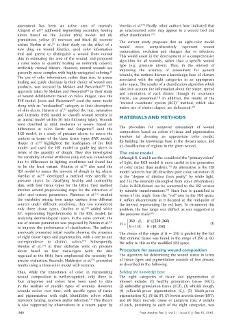
involves several preprocessing steps for the extraction of by suitable transformations. Since hue is quantified in
[18]
color and texture parameters. Wannous et al. studied terms of the angle from the “red”‑axis in the HSI space,
[11]
the variability arising from image capture from different it suffers discontinuity at 0 (located at the mid‑point of
sources under different conditions, they too considered the interval representing the red hue). To circumvent this
only three tissue types. Dorileo et al. added white problem, the hue range was shifted, as was suggested in
[12]
(W, representing hyperkeratosis) to the RYK model, for the previous study. [20]
analyzing dermatological ulcers. In the same context, the
360
[13]
use of texture parameters was proposed by Pereira et al. H = − H ∈ H, [250, 360) (1)
to improve the performance of classification. The authors H + H ∈110, [0, 250)
previously presented initial results showing the presence The choice of the origin at H = 250 is guided by the fact
of eight tissue types and pigmentation, with a one‑to‑one that minimal tissue was found in the range of 250 ± 60.
correspondence to distinct colors. Subsequently, We refer to this as the modified HSI space.
[14]
Veredas et al., in their elaborate work on pressure
[15]
ulcers based on four tissue‑types (with the skin Procedure for assessing wound composition
regarded as the fifth), have emphasized the necessity for The algorithm for determining the wound status in terms
precise evaluation. Recently, Mukherjee et al. presented of tissue types and pigmentation consists of two phases,
[16]
results using a three‑color model with textures. as described in the following.
Thus, while the importance of color in representing Building the knowledge base
wound composition is well‑recognized, only three to The eight categories of tissue and pigmentation of
four categories and colors have been used to date interest include: (1) healthy granulation tissue (HGT),
in the analysis of specific types of wounds. However, (2) unhealthy granulation tissue (UGT), (3) whitish slough,
wounds evolve over time, with specific types of tissue (4) yellowish‑green pigmentation (G ), (5) bluish‑green
1
and pigmentation with eight identifiable colors which pigmentation (G ), (6) fat (F), (7) brown necrotic tissue (BNT),
2
represent healing, necrosis and/or infection. This theory and (8) black necrotic tissue or gangrene (Ga). A sample
[14]
is also supported by observations in a recent paper by of each, pertaining to each of the eight categories, was
262 Plast Aesthet Res || Vol 2 || Issue 5 || Sep 15, 2015

