
Page 127 - Read Online
P. 127
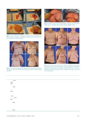
Figure 2: Relying on preoperative topographic markings, fat was injected
                                                              on the breast in multiple tissue planes, through multiple passes


          Figure  1:  Fat is allowed to separate by gravity and then refined on a
          Tefla Pad until it reached a custard-like consistency. The refined fat was
          then transferred in 10-mL syringes


                                                              Figure  4:  A  62-year-old female with a history of bilateral mastectomy
                                                              and radiation therapy on the left breast; followed bilateral deep inferior
          Figure 3: A 52-year-old female who had a left lumpectomy and radiation   epigastric perforator flap reconstruction. She benefited from 2 rounds of
          3  years ago. She was treated with lipoinjection of the lateral contour   fat  injection  of the  left  upper breast  contracture  and serial  excision  of
          deformity                                           breast skin paddle


          Table 1: Review of the literature
           Authors         Year  Number of  Average volume of                  Complications
                                patients (n)  fat injection (mL)
           Pérez-Cano et al. [20]  2012  71      140       14.1% of patients developed cysts
           Khouri et al. [21]  2012  81          277       16% of patients report fat necrosis after 1-year
           Rubin et al. [22]  2012  27          526.5      25.5% of patients developed oil cysts
                                                           17.1% of patients developed fat necrosis
           De Blacam et al. [23]  2011  49       67        3.6% of patients developed fat necrosis
                                                           1.8% of patients developed oil cysts
                                                           0.9% of patients developed infections
           Kijima et al. [24]  2012  21          123       4.7% of patients developed fat necrosis
                                                           4.7% of patients developed infection
           Kamakura and Ito [25]  2011  20       240       11% of patients developed oil cysts
           Losken et al. [26]  2011  107         40        11% of patients reported fat necrosis, erythema, keloid scarring, and pain
           Serra-Renom et al. [27]  2011  28    39.36      0% fat stable in all patients
           Sinna et al. [28]  2010  244          176       2% of patients developed fat necrosis
                                                           1.2% of patients developed infection
           Yoshimura et al. [29]  2010  15       264       0% no reported complications
           Illouz and      2009    820           145       9.2% of patients developed bruising
           Sterodimas [30]                                 4.3% of patients developed striae
                                                           1.4% of patients developed hematomas
                                                           0.6% of patients developed infections
           Panettiere et al. [31]  2009  61      24.5      0% no reported complications


          Plast Aesthet Res || Vol 2 || Issue 3 || May 15, 2015                                             117