
Page 83 - Read Online
P. 83
At the end of follow‑up dimensional reduction, the WBS Six out of 16 (38%) chronic ulcers healed in a mean
and presence of the adverse reaction were recorded. time of 42 ± 16 days. The application of the new skin
substitutes reduced the mean percentage of the wound
The primary endpoint of the study was to determine the dimension by 70% (P = 0.0007). The WBS demonstrated
variation in ulcer dimension versus t0. Secondary endpoints an improvement of 52% (P < 0.0001) at the end of the
included the evaluation of variation in the wound bed and follow‑up period from the score recorded at the entry of
exudate to determine the WBS, the percentage of wound the study.
reduction, and the percentage and time to healing.
Case example: ankle diabetic ulcers in a 66‑year‑old
Abstracted data were stored using an Excel Office patient treated with allogeneic keratinocyte [Figures 1–5].
database (Microsoft Corporation, Washington, USA)
containing fields for clinical data entry. The statistical DISCUSSION
analysis was performed considering the patient as a
unit of analysis initially (for anamnestic data), and then The chronic wound microenvironment is biologically
the single‑chronic wound (for clinical results). The mean distinct from the acute wound milieu: venous and diabetic
reduction of the skin lesion during follow‑up was verified chronic ulcers are hypothesized to be trapped in the
with the Wilcoxon Signed‑Rank Test. The variation of WBS inflammatory and proliferative phases of normal healing,
versus t0 was analyzed with the Friedman test. The level respectively. Poor wound healing may be a consequence
of statistical significance was fixed to 5% (P < 0.05) to of abnormal insulin signaling and hyperglycemia, affecting
reject the null hypothesis.
skin proliferation and differentiation. Skin biopsies
[10]
performed in nondiabetic and diabetic subjects from
RESULTS the edges of chronic wounds have revealed increased
expression of transforming growth factor (TGF) beta 3 and
Between January of 2011 and December of 2013, low expression of TGF‑beta 1, resulting in nonhealing.
[11]
11 patients with diabetes type 2 with 16 wounds Abnormal expression of insulin‑like growth factor type 1
underwent an application of allogeneic epidermal in diabetic skin may also contribute to delayed wound
substitutes on a hyaluronic acid scaffold. Table 1 healing. [12]
describes the demographics of all patients. Four out of
11 (36%) patients were females, and the mean age of The damaged biological background of diabetic wounds
all patients was 75 ± 8.2 years. Among comorbidities, explains the necessity to modulate therapeutically the
hypertension (7, 63%) and cardiopathy (3, 27%) were the unbalanced levels of growth factors, signaling molecules,
[13]
most frequent. and extracellular matrix proteins. How these novel
skin substitutes work is still not completely understood.
As described by Table 2, 8 out of 16 (50%) chronic ulcers Initially, especially with skin substitutes as cultured
were located on the ankle or foot, and the other 50% were epidermis and living bilayered skin construct, some
located on the lower third of the leg. Five (32%) ulcers degree of permanent engraftment was thought to assist
showed a mild to moderate local infection at the entry of in healing. As shown by DNA and Y chromosome probes,
the study; only 1 severe infection was present. The mean allogeneic constructs are not the same as autografts: a true
preoperative wound size for all diabetic chronic ulcers take of allogeneic sheets has not been demonstrated.
[14]
was 14.37 ± 9.29 cm , and the mean preoperative WBS The allogeneic skin constructs usually do not stay on the
2
was 9.6 ± 3.5. During the follow‑up period, no wound wound for more than a few weeks, and their function is
advanced to require amputation of the foot or lower limb, not to replace tissues or cells, but instead to stimulate
and only one ulcer developed a local severe infection tissue repair as pharmacologic agents, secreting healing
under the application of the sheet, enlarging the wound factors in chronic wound microenvironment. The key role
dimensions. No other adverse reactions were recorded. of allogeneic constructs seems to be the secretion of
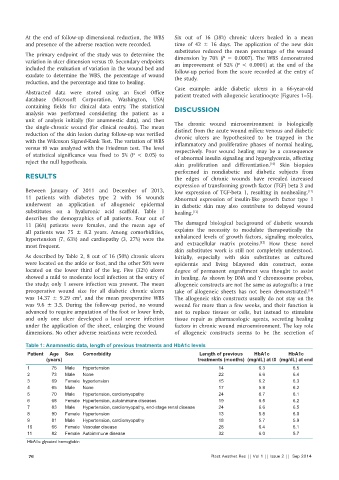
Table 1: Anamnestic data, length of previous treatments and HbA1c levels
Patient Age Sex Comorbidity Length of previous HbA1c HbA1c
(years) treatments (months) (mg/dL) at t0 (mg/dL) at end
1 75 Male Hypertension 14 6.3 6.5
2 73 Male None 22 6.6 6.4
3 69 Female hypertension 15 6.2 6.3
4 65 Male None 17 5.9 6.2
5 70 Male Hypertension, cardiomyopathy 24 6.7 6.1
6 68 Female Hypertension, autoimmune diseases 19 6.5 6.2
7 83 Male Hypertension, cardiomyopathy, end-stage renal disease 24 6.6 6.5
8 90 Female Hypertension 13 5.8 6.0
9 81 Male Hypertension, cardiomyopathy 18 5.7 5.9
10 66 Female Vascular disease 28 6.4 6.1
11 82 Female Autoimmune disease 32 6.0 5.7
HbA1c: glycated hemoglobin
76 Plast Aesthet Res || Vol 1 || Issue 2 || Sep 2014