
Page 44 - Read Online
P. 44
at the level of the shaft of the proximal phalanx of the artery of the thumb, facilitating end-to-end repair without
thumb [Figure 4]. Intraoperatively, transection of the ulnar a vein graft. The digital vein of the index finger was also
digital artery was at the level of the base of the thumb. utilized to allow venous drainage of the thumb [Figure 6].
The thumb was ischemic. Skeletal fixation was achieved On release of the tourniquet, there was good blood flow
using Kirschner wires [Figure 5]. Dissection of the digital into the thumb and fingers [Figure 7]. Postoperatively,
nerve revealed contused nerve fibers, although the fibers the patient received six sessions of hyperbaric oxygen
were intact. The ulnar digital artery was found to be therapy. After an uneventful hospital course, the patient
transected at the shaft level with segmental loss; this was discharged on the tenth postoperative day.
segmental loss made primary repair difficult. The radial
digital artery and the nerve of the thumb were contused DISCUSSION
but intact. The dorsal veins were contused.
To bridge the gap, the superficial palmar arch was Avulsion injuries to the thumb can result in extensive
dissected until its ulnar end where the branch to the damage to long segments of vessels, which makes direct
common digital artery to the index and middle fingers suturing of the structures difficult. The decision to proceed
arising from the palmar arch. A digital clamp was placed with revascularizing an avulsed thumb depends on several
over the ulnar end of the palmar arch and the radial digital factors, including the mechanism of injury, the patient’s
artery of the index finger. The tourniquet was released age, occupation and hand dominance, and overall medical
and the vascularity of the index finger was confirmed, condition and intraoperative assessment of the injured
before reapplying the tourniquet. A Vascular clip was structures. When the decision is taken to retain the thumb
used to ligate the superficial palmar arch just before the by revascularization, the options available are to use a
division of common digital artery to the index and middle vein graft to reconstruct the segmental loss or to transfer
fingers. To obtain additional length, the radial digital nearby vessels to adequately bridge the gap of the injured
artery to the index finger was also ligated and divided.
The palmar arch was turned to reach the ulnar digital
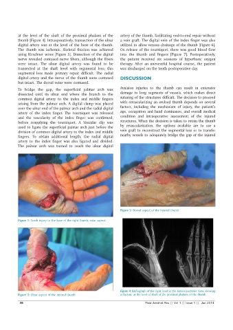
Figure 2: Dorsal aspect of the injured thumb
Figure 1: Crush injury to the base of the right thumb, volar aspect
Figure 4: Radiograph of the right hand in the antero-posterior view, showing
Figure 3: Ulnar aspect of the injured thumb a fracture at the level of shaft of the proximal phalanx of the thumb
38 Plast Aesthet Res || Vol 1 || Issue 1 || Jun 2014