
Page 40 - Read Online
P. 40
the superficial temporal artery with Onyx Liquid Embolic this would be an easy procedure to do as a second stage
System (Onyx HD-500) [Figure 6]. [4,5] This resulted in should the pinna component become problematic.
®
reduction in lesion size and its vascularity. The planned There were no complications related to either the
excision included the skin directly overlying the parotid as preoperative angiography or embolization procedure.
well as the lower half of the ear [Figure 7] and extended The patient was discharged on day 5 after the procedure.
down into the neck, to allow for closure of the defect as a Histology confirmed arteriovenous malformation involving
cervicofacial rotation advancement flap. Careful dissection the subcutaneous tissue and parotid gland without
allowed for retrograde identification of the facial nerve any atypia or malignancy present. There has been no
branches. The tumor was circumscribed and simultaneous recurrence to our knowledge so far.
dissection performed in all directions [Figure 8]. It was
possible through this approach to then remove the entire DISCUSSION
tumor superficial and deep to the facial nerve, including
the lower part of the ear [Figures 9 and 10]. It was decided Maxillofacial VMs are formed due to an error of vascular
at the end of the procedure not to remove the remaining morphogenesis. They may correspond to a defective
components of the pinna as these are quite asymptomatic remodeling process at the final stages of vessel formation.
and removing them wound mean probably having to use a Although no hereditary VM exist, the defect might be
temporal parietal fascia and covering it with a skin graft, genetically based and secondarily expressed in the first
which is considered unnecessary at present. However, few years of life. VM generally grow in proportion to the
growth of the affected child, but may increase in size
secondary to various triggering factors such as increased
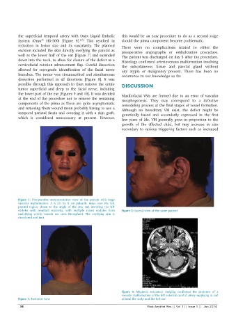
Figure 1: Preoperative anteroposterior view of the patient with large
vascular malformation. A 6 cm by 8 cm pulsatile mass over the left
parotid region, down to the angle of the jaw, and involving the left
earlobe with resultant macrotia, with multiple raised nodules form Figure 2: Lateral view of the same patient
underlying ectatic vessels are seen throughout. The overlying skin is
discolored and taut
Figure 4: Magnetic resonance imaging confirmed the presence of a
vascular malformation of the left external carotid artery supplying in and
Figure 3: Posterior view around the scalp and the left ear
34 Plast Aesthet Res || Vol 1 || Issue 1 || Jun 2014