
Page 41 - Read Online
P. 41
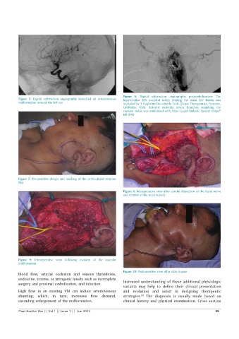
Figure 6:  Digital  subtraction angiography postembolization:  The
          Figure 5:  Digital  subtraction  angiography  identified  an  arteriovenous   hypertrophic left occipital artery feeding  the  main  A-V  fistula was
          malformation around the left ear                    occluded by 3 Guglielmi Detachable Coils (Target Therapeutics, Fremont,
                                                              California,  USA). Anterior  auricular artery  branches supplying the
                                                              vascular nidus was embolized with Onyx Liquid Embolic System (Onyx
                                                                                                              ®
                                                              HD-500)


          Figure 7:  Preoperative design and marking of the cervicofacial rotation
          flap

                                                              Figure 8: Intraoperative view after careful dissection of the facial nerve
                                                              and control of the neck vessels


          Figure 9:  Intraoperative  view  following excision  of the  vascular
          malformation

                                                              Figure 10: Postoperative view after skin closure
          blood  flow,  arterial  occlusion  and  venous  thrombosis,
          endocrine, trauma, or iatrogenic insults such as incomplete   Increased understanding of these additional physiologic
          surgery and proximal embolization, and infection.
                                                              variants may help to define their clinical presentation
          High  flow in an existing  VM can induce arteriovenous   and evolution and assist in designing therapeutic
          shunting,  which,  in  turn,  increases  flow demand,   strategies.  The diagnosis is usually made based on
                                                                       [6]
          cascading enlargement of the malformation.          clinical  history  and  physical  examination.  Cross  section

          Plast Aesthet Res || Vol 1 || Issue 1 ||  Jun 2014                                                35