
Page 85 - Read Online
P. 85
Kobylarz et al. Plast Aesthet Res 2023;10:2 https://dx.doi.org/10.20517/2347-9264.2022.38 Page 9 of 20
Figure 5. EMG recording demonstrating EMG activity with a superimposed artifact in the masseter, orbicularis oculi, and orbicularis oris
muscles during the surgical procedure. Amplitude is 20 µV per division. The sweep speed is 400 msec per division.
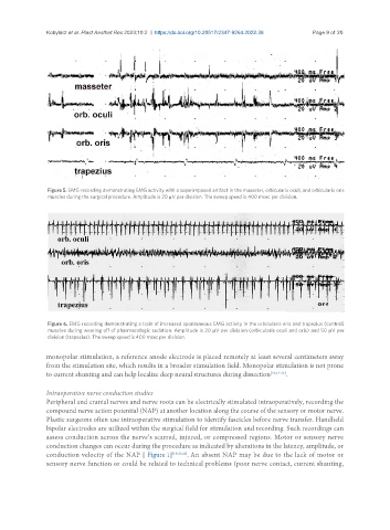
Figure 6. EMG recording demonstrating a train of increased spontaneous EMG activity in the orbicularis oris and trapezius (control)
muscles during wearing off of pharmacologic sedation. Amplitude is 20 µV per division (orbicularis oculi and oris) and 50 µV per
division (trapezius). The sweep speed is 400 msec per division.
monopolar stimulation, a reference anode electrode is placed remotely at least several centimeters away
from the stimulation site, which results in a broader stimulation field. Monopolar stimulation is not prone
to current shunting and can help localize deep neural structures during dissection [18,21-23] .
Intraoperative nerve conduction studies
Peripheral and cranial nerves and nerve roots can be electrically stimulated intraoperatively, recording the
compound nerve action potential (NAP) at another location along the course of the sensory or motor nerve.
Plastic surgeons often use intraoperative stimulation to identify fascicles before nerve transfer. Handheld
bipolar electrodes are utilized within the surgical field for stimulation and recording. Such recordings can
assess conduction across the nerve’s scarred, injured, or compressed regions. Motor or sensory nerve
conduction changes can occur during the procedure as indicated by alterations in the latency, amplitude, or
conduction velocity of the NAP [ Figure 1] [18,20,22] . An absent NAP may be due to the lack of motor or
sensory nerve function or could be related to technical problems (poor nerve contact, current shunting,