
Page 82 - Read Online
P. 82
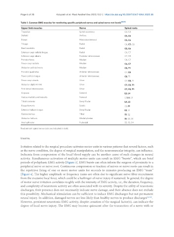
Page 6 of 20             Kobylarz et al. Plast Aesthet Res 2023;10:2  https://dx.doi.org/10.20517/2347-9264.2022.38

               Table 1. Common EMG muscles for monitoring specific peripheral nerves and spinal nerve root levels [16,17]
                Upper limb muscles                     Nerve                          Spinal roots
                Trapezius                              Spinal accessory               C3, C4
                Deltoid                                Axillary                       C5, C6
                Biceps                                 Musculocutaneous               C5, C6
                Triceps                                Radial                         C6, C7, C8
                Brachioradialis                        Radial                         C5, C6
                Extensor carpi radialis longus         Radial                         C6, C7
                Extensor carpi ulnaris                 Posterior interosseous         C7, C8
                Pronator teres                         Median                         C6, C7
                Flexor carpi radialis                  Median                         C6, C7
                Abductor pollicis brevis               Median                         C8, T1
                Pronator quadratus                     Anterior interosseous          C7, C8
                Flexor pollicis longus                 Anterior interosseous          C8, T1
                Flexor carpi ulnaris                   Ulnar                          C7, C8, T1
                Abductor digiti minimi                 Ulnar                          C7, C8, T1
                First dorsal interosseous              Ulnar                          C7, C8, T1
                Iliopsoas                              Femoral                        L3, L4
                Vastus medialis and lateralis          Femoral                        L3,L4, L5
                Tibialis anterior                      Deep fibular                   L4, L5
                Biceps femoris                         Sciatic                        L5, S1
                Extensor hallucis longus               Deep fibular                   L5, S1
                Gastrocnemius                          Tibial                         S1, S2
                Abductor hallucis                      Medial plantar                 S1, S2, S3
                Anal sphincter                         Pudendal                       S2, S3, S4
               Predominant spinal nerve roots are indicated in bold.


               trauma.


               Irritation related to the surgical procedure activates motor units in various patterns that several factors, such
               as the nerve condition, the degree of surgical manipulation, and the neuromuscular integrity, can influence.
               Ischemia from compression of the local blood supply can be another cause of such changes in neural
               activity. Simultaneous activation of multiple motor units can result in EMG “bursts”, which are brief
               periods of polyphasic EMG activity [Figure 3]. EMG bursts can often inform the surgeon of proximity to a
               peripheral nerve or nerve root. Continuous compression or traction of nerves or nerve roots can result in
               the repetitive firing of one or more motor units for seconds to minutes producing an EMG “train”
               [Figure 4]. The higher amplitude or frequency trains are often due to significant nerve fiber recruitment
               from the excessive local force, which could be a harbinger of nerve injury if sustained. In general, the degree
               of motor nerve irritation correlates roughly with the intensity of EMG activity, i.e., the duration, frequency,
               and complexity of neurotonic activity are often associated with its severity. Despite the utility of neurotonic
               discharges, their presence does not necessarily indicate nerve damage, and their absence does not exclude
               this possibility. Mechanical stimulation can be sufficient to induce EMG discharges but not permanent
               neural injury. In addition, damaged nerves are less likely than healthy nerves to produce discharges [18-20] .
               However, persistent neurotonic EMG activity, despite cessation of the surgical factor(s), can indicate the
               degree of local nerve injury. The EMG may become quiescent after the transection of a nerve with or