
Page 86 - Read Online
P. 86
Page 10 of 20 Kobylarz et al. Plast Aesthet Res 2023;10:2 https://dx.doi.org/10.20517/2347-9264.2022.38
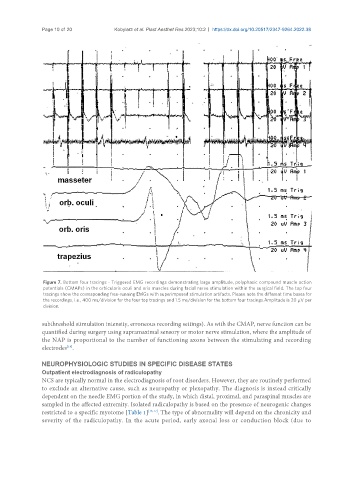
Figure 7. Bottom four tracings - Triggered EMG recordings demonstrating large amplitude, polyphasic compound muscle action
potentials (CMAPs) in the orbicularis oculi and oris muscles during facial nerve stimulation within the surgical field. The top four
tracings show the corresponding free-running EMGs with superimposed stimulation artifacts. Please note the different time bases for
the recordings, i.e., 400 ms/division for the four top tracings and 1.5 ms/division for the bottom four tracings.Amplitude is 20 µV per
division.
subthreshold stimulation intensity, erroneous recording settings). As with the CMAP, nerve function can be
quantified during surgery using supramaximal sensory or motor nerve stimulation, where the amplitude of
the NAP is proportional to the number of functioning axons between the stimulating and recording
electrodes .
[19]
NEUROPHYSIOLOGIC STUDIES IN SPECIFIC DISEASE STATES
Outpatient electrodiagnosis of radiculopathy
NCS are typically normal in the electrodiagnosis of root disorders. However, they are routinely performed
to exclude an alternative cause, such as neuropathy or plexopathy. The diagnosis is instead critically
dependent on the needle EMG portion of the study, in which distal, proximal, and paraspinal muscles are
sampled in the affected extremity. Isolated radiculopathy is based on the presence of neurogenic changes
restricted to a specific myotome [Table 1] [16,17] . The type of abnormality will depend on the chronicity and
severity of the radiculopathy. In the acute period, early axonal loss or conduction block (due to