
Page 89 - Read Online
P. 89
Kobylarz et al. Plast Aesthet Res 2023;10:2 https://dx.doi.org/10.20517/2347-9264.2022.38 Page 13 of 20
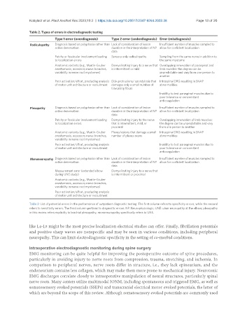
Table 2. Types of errors in electrodiagnostic testing
Type 1 error (overdiagnosis) Type 2 error (underdiagnosis) Error (misdiagnosis)
Radiculopathy Diagnosis based on polyphasia rather than Lack of consideration of lesion Insufficient number of muscles sampled to
active denervation duration in the interpretation of NP allow for confident localization
data
Patchy or fascicular involvement leading Sensory only radiculopathy Sampling from the same nerve in addition to
to localization errors the same myotome
Anatomic variants (e.g., Martin-Gruber Demyelinating injury to a nerve that Overlapping innervation of paraspinal and
anastomosis, accessory nerve branches, is intermittent or mild limb muscles: the degree can be
variability in nerve root myotomes) unpredictable and vary from one person to
another
Poor activation/effort, precluding analysis Disk protrusions/ spondylosis that Intraspinal DRG resulting in SNAP
of motor unit architecture or recruitment damages only a small number of abnormalities
traversing fibers
Inability to test paraspinal muscles due to
poor tolerance or concomitant
anticoagulation
Plexopathy Diagnosis based on polyphasia rather than Lack of consideration of lesion Insufficient number of muscles sampled to
active denervation duration in the interpretation of NP allow for confident localization
data
Patchy or fascicular involvement leading Demyelinating injury to the nerve Overlapping innervation of limb muscles:
to localization errors that is intermittent, mild, or the degree can be unpredictable and vary
proximal from one person to another
Anatomic variants (e.g., Martin-Gruber Plexus lesions that damage a small Intraspinal DRG resulting in SNAP
anastomosis, accessory nerve branches, number of plexus axons abnormalities
variability in nerve root myotomes)
Poor activation/effort, precluding analysis Inability to test paraspinal muscles due to
of motor unit architecture or recruitment poor tolerance or concomitant
anticoagulation
Mononeuropathy Diagnosis based on polyphasia rather than Lack of consideration of lesion Insufficient number of muscles sampled to
active denervation duration in the interpretation of NP allow for confident localization
data
Measurement error (extended elbow Demyelinating injury to a nerve that
during UNE study) is intermittent or proximal
Anatomic variants (e.g., Martin-Gruber
anastomosis, accessory nerve branches,
variability in nerve root myotomes)
Poor activation/effort, precluding analysis
of motor unit architecture or recruitment
Table 2: List of potential errors in the performance of outpatient diagnostic testing. The first column refers to specificity errors, while the second
refers to sensitivity errors. The third column pertains to diagnostic errors. NP: Neurophysiologic; UNE: ulnar neuropathy at the elbow; plexopathy
in this review refers explicitly to brachial plexopathy; mononeuropathy specifically refers to UNE.
like L4-L5 might be the most precise localization electrical studies can offer. Finally, fibrillation potentials
and positive sharp waves are nonspecific and may be seen in various conditions, including peripheral
neuropathy. This can limit electrodiagnostic specificity in the setting of co-morbid conditions.
Intraoperative electrodiagnostic monitoring during spine surgery
EMG monitoring can be quite helpful for improving the postoperative outcome of spine procedures,
particularly in avoiding injury to nerve roots from compression, trauma, stretching, and ischemia. In
comparison to peripheral nerves, nerve roots differ in structure, i.e., they lack epineurium, and the
endoneurium contains less collagen, which may make them more prone to mechanical injury. Neurotonic
EMG discharges correlate closely to intraoperative manipulation of neural structures, particularly spinal
nerve roots. Many centers utilize multimodal IONM, including spontaneous and triggered EMG, as well as
somatosensory evoked potentials (SSEPs) and transcranial electrical motor evoked potentials, the latter of
which are beyond the scope of this review. Although somatosensory evoked potentials are commonly used