
Page 81 - Read Online
P. 81
Kobylarz et al. Plast Aesthet Res 2023;10:2 https://dx.doi.org/10.20517/2347-9264.2022.38 Page 5 of 20
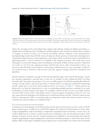
Figure 2. Motor unit action potential. Amplitude reflects the number of muscle fibers contributing to a potential elicited from a single
nerve fiber. Duration reflects dispersion in neuromuscular activation of branches to muscle fibers. Phases reflect baseline crossings and
relate to the increased complexity of motor units due to reinnervation. Amplitude is 100 µV per division. The sweep speed is 5 msec per
division.
within the operating room, neurophysiologic signals must undergo analog and digital processing, i.e.,
amplification and filtering. The recordings are visually displayed and can also be broadcast from a speaker
so changes in muscle activity can be heard, providing auditory feedback to the intraoperative
neurophysiologic monitoring (IONM) team and surgeons. Bursts and runs of neurotonic discharges have
characteristic high-frequency visual appearances and sounds, which can alert the team of perturbation or
impending injury to neural structures if correlated to the surgical procedure. We would expect such
discharges to occur in the setting of nerve perturbation. Alterations of EMG activity and nerve conduction
can result not only from the surgical procedure itself but also due to other intraoperative factors, e.g.,
neuromuscular blockade, level of sedation, temperature, ambient electrical noise, neurologic conditions,
and technical issues. Therefore, false positive and false negative changes should be considered and
identified.
Muscle selection to maximize coverage for the relevant anatomic region is the best IONM strategy. Careful
pre-operative planning is essential since it may not be possible to place additional EMG recording
electrodes after the surgery is in progress. The most accessible muscles for IONM lie superficially within the
cranial, cervical, lumbar, and sacral myotomes. Common EMG recording sites utilized for monitoring
specific spinal nerve root levels and peripheral nerves are shown in Table 1 [16,17] . Most muscles are
innervated by at least two adjacent nerve roots, so monitoring multiple muscles is essential for accurate
localization of neural irritation and injury. For example, irritation of the L4 nerve root might result in
abnormal EMG activity solely in the tibialis anterior, whereas surgical manipulation of the L5 nerve root
may cause abnormal discharges in the tibialis anterior and biceps femoris muscles. Technical issues can also
affect the intraoperative EMG, so simultaneously recording from uninvolved remote muscles to serve as
controls can be of great utility, e.g., troubleshooting .
[3-7]
Free-running spontaneous electromyography
Continuous electromyography is monitored from muscles innervated by nerves or nerve roots at risk for
injury during surgery. Usually, the recorded EMG signals will consist of a broad mixture of motor units.
Surgical manipulation, such as stretching or compression of peripheral nerves, branches, or nerve roots, can
result in high-frequency bursts or trains of neurotonic discharges in the corresponding muscle(s). Such
changes in muscle activity provide valuable instantaneous feedback to the monitoring team and the
surgeons regarding the surgical manipulation and actions and their proximity to neural structures. In this
manner, EMG monitoring is beneficial for preventing severe or irreversible nerve injury from inadvertent