
Page 79 - Read Online
P. 79
Kobylarz et al. Plast Aesthet Res 2023;10:2 https://dx.doi.org/10.20517/2347-9264.2022.38 Page 3 of 20
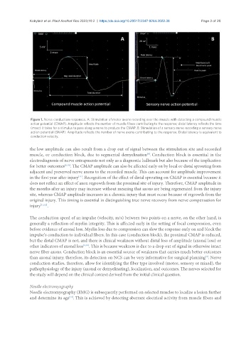
Figure 1. Nerve conduction responses. A: Stimulation of motor axons recording over the muscle with detecting a compound muscle
action potential (CMAP). Amplitude reflects the number of muscle fibers contributing to the response; distal latency reflects the time
(msec) it takes for a stimulus to pass along a nerve to produce the CMAP. B: Stimulation of a sensory nerve recording a sensory nerve
action potential (SNAP). Amplitude reflects the number of nerve axons contributing to the response. Distal latency is equivalent to
conduction velocity.
the low amplitude can also result from a drop out of signal between the stimulation site and recorded
muscle, or conduction block, due to segmental demyelination . Conduction block is essential in the
[8]
electrodiagnosis of nerve entrapments not only as a diagnostic hallmark but also because of the implication
for better outcomes [9,10] . The CMAP amplitude can also be affected early on by local or distal sprouting from
adjacent and preserved nerve axons to the recorded muscle. This can account for amplitude improvement
in the first year after injury . Recognition of the effect of distal sprouting on CMAP is essential because it
[11]
does not reflect an effect of axon regrowth from the proximal site of injury. Therefore, CMAP amplitude in
the months after an injury may increase without meaning that axons are being regenerated from the injury
site, whereas CMAP amplitude increases in a chronic injury that must occur because of regrowth from the
original injury. This timing is essential in distinguishing true nerve recovery from nerve compensation for
injury [11,12] .
The conduction speed of an impulse (velocity, m/s) between two points on a nerve, on the other hand, is
generally a reflection of myelin integrity. This is affected early in the setting of focal compression, even
before evidence of axonal loss. Myelin loss due to compression can slow the response early on and block the
impulse’s conduction to individual fibers. In this case (conduction block), the proximal CMAP is reduced,
but the distal CMAP is not, and there is clinical weakness without distal loss of amplitude (axonal loss) or
other indicators of axonal loss [9,10] . This is because weakness is due to a drop out of signal in otherwise intact
nerve fiber axons. Conduction block is an essential source of weakness that carries much better outcomes
than axonal injury; therefore, its detection on NCS can be very informative for surgical planning . Nerve
[4]
conduction studies, therefore, allow for identifying the fiber type involved (motor, sensory or mixed), the
pathophysiology of the injury (axonal or demyelinating), localization, and outcomes. The nerves selected for
the study will depend on the clinical context derived from the initial clinical question.
Needle electromyography
Needle electromyography (EMG) is subsequently performed on selected muscles to localize a lesion further
and determine its age . This is achieved by detecting aberrant electrical activity from muscle fibers and
[13]