
Page 28 - Read Online
P. 28
Won et al. Plast Aesthet Res 2019;6:6 I http://dx.doi.org/10.20517/2347-9264.2018.82 Page 9 of 16
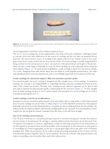
Figure 11. Perichondrium on the undersurface of the radix portion of the graft with slight rasping of the bone can reduce mobility and
decrease the chances of migration
Dorsal augmentation with diced costal cartilage wrapped in fascia
The use of diced cartilage for dorsal augmentation has been previously published. Although similar
in concept, there have been differences in the source of cartilage and the way they are prepared before
insertion. The most common source of cartilage is the septum and ear if the needed volume is not small.
Superiority of one versus another has not been clearly shown. The diced cartilage is usually wrapped before
being inserted into the dorsal pocket so that it can conform to the desired shape and height of the dorsum.
There are also a wide range of materials to wrap the diced cartilage in, with temporalis fascia being the
most popular [Figure 14]. To avoid dorsal irregularities, costal cartilage is diced into fine pieces (usually
0.5-1 mm). Wrapping them with thicker fascia lata also helps to avoid palpable protuberances. The radix
and supratip portions need extra attention in order to avoid slight depression in the long-term follow up.
Costal cartilage for structural support: SEG and extended spreader grafts
For structural grafts, the costal cartilage is designed into flat, straight pieces of thin cartilage. To minimize
warping, the cartilage is cut longitudinally or tangentially, leaving symmetric peripheral portions on both
sides of the central core portion. However, when warping occurs, affected fragments can be split in half
and used as bilateral extended spreader grafts, compensating for the curvature [Figure 15]. If a thin straight
piece of septal cartilage remains, it can be used as septal extension graft and costal cartilage can be used as
extended spreader grafts to fix it.
Costal cartilage used for tip modifications
Examples are lateral crural strut grafts, lateral crural onlay grafts and tip onlay grafts. Usually thin, beveled
slices of costal cartilage are carved with a 10 blade [Figure 16]. Grafts should be symmetrical when applied
bilaterally and not thick in thin skinned patients. Cap graft can also be fashioned from the rib cartilage. The
authors prefer an elliptical shaped, well beveled cartilage graft for the cap graft. The perichondrium can be
draped over the cap graft for smooth transition from the dome to the soft tissue triangle.
Use of rib cartilage perichondrium
The rib perichondrium is a valuable grafting material in revision rhinoplasty. Usually the anterior
perichondrium of the harvested rib cartilage is used but additional perichondrium can be harvested from
the adjacent ribs (superior or inferior). Applications include the following: placement in the undersurface of
the dorsal graft to increase friction and avoid mobility of the dorsal graft, on the dorsum for a radix graft or
to camouflage any dorsal irregularities, on the tip to conceal graft edges and in any other areas of thinned/
damaged skin. When harvesting the rib, the authors prefer harvesting periosteum of the rib bone near the
costochondral junction together with the perichondrium. Periosteum is thicker than perichondrium and
helps to camouflage more especially when used at the radix portion.