
Page 25 - Read Online
P. 25
Page 6 of 16 Won et al. Plast Aesthet Res 2019;6:6 I http://dx.doi.org/10.20517/2347-9264.2018.82
A B
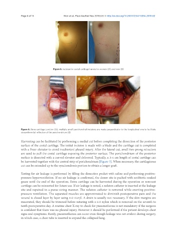
Figure 5. Incision for costal cartilage harvest in women (A) and men (B)
A B
Figure 6. Bone cartilage junction (A); multiple small perichondrial incisions are made perpendicular to the longitudinal one to facilitate
circumferential reflection of the perichondrium (B)
Harvesting can be facilitated by performing a medial cut before completing the dissection of the posterior
surface of the costal cartilage. The initial incision is made with a blade and the cartilage cut is completed
with a Freer elevator to avoid inadvertent pleural injury. After the lateral cut, small two-prong retractors
are used to pull the costal cartilage exposing the posterior surface. The perichondrium of the posterior
surface is dissected with a curved elevator and delivered. Typically, a 3-4 cm length of costal cartilage can
be harvested together with the central strip of perichondrium [Figure 7]. When necessary, the cartilaginous
cut can be extended up to the synchondrosis portion to obtain a longer graft.
Testing for air leakage is performed by filling the dissection pocket with saline and performing positive-
pressure hyperventilation. If no air leakage is confirmed, the donor site is packed with antibiotic-soaked
gauze until the end of the operation. Extra cartilage can be harvested during the operation or remnant
cartilage can be reinserted for future use. If air leakage is noted, a nelaton catheter is inserted at the leakage
site and repaired in a purse-string manner. The nelaton catheter is removed while exerting positive-
pressure ventilation. The separated muscles are approximated to diminish postoperative pain and the
wound is closed layer by layer using 4-0 vicryl. A drain is usually not necessary. If the skin margins are
macerated, they should be trimmed before suturing with a 6-0 nylon which is removed on the seventh to
tenth postoperative day. A routine chest X-ray to check for pneumothorax is not mandatory if the surgeon
is confident that there was no pleural injury. However it should be performed if the patient develops chest
signs and symptoms. Rarely, pneumothorax can occur even though leakage was not evident during surgery,
in which case, a chest tube is inserted to expand the collapsed lung.