
Page 26 - Read Online
P. 26
Won et al. Plast Aesthet Res 2019;6:6 I http://dx.doi.org/10.20517/2347-9264.2018.82 Page 7 of 16
Figure 7. Harvested costal cartilage together with the central strip of perichondrium
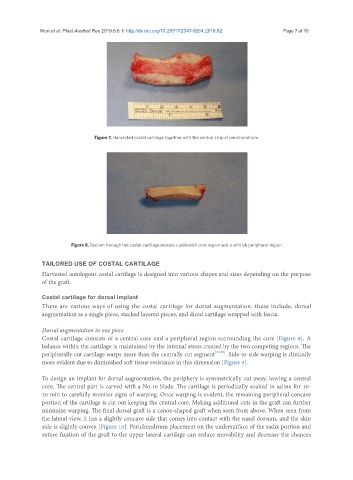
Figure 8. Section through the costal cartilage reveals a yellowish core region and a whitish peripheral region
TAILORED USE OF COSTAL CARTILAGE
Harvested autologous costal cartilage is designed into various shapes and sizes depending on the purpose
of the graft.
Costal cartilage for dorsal implant
There are various ways of using the costal cartilage for dorsal augmentation, these include, dorsal
augmentation as a single piece, stacked layered pieces, and diced cartilage wrapped with fascia.
Dorsal augmentation in one piece
Costal cartilage consists of a central core and a peripheral region surrounding the core [Figure 8]. A
balance within the cartilage is maintained by the internal stress created by the two competing regions. The
peripherally cut cartilage warps more than the centrally cut segment [21,22] . Side-to-side warping is clinically
more evident due to diminished soft tissue resistance in this dimension [Figure 9].
To design an implant for dorsal augmentation, the periphery is symmetrically cut away, leaving a central
core. The central part is carved with a No.10 blade. The cartilage is periodically soaked in saline for 10-
20 min to carefully monitor signs of warping. Once warping is evident, the remaining peripheral concave
portion of the cartilage is cut out keeping the central core. Making additional cuts in the graft can further
minimize warping. The final dorsal graft is a canoe-shaped graft when seen from above. When seen from
the lateral view, it has a slightly concave side that comes into contact with the nasal dorsum, and the skin
side is slightly convex [Figure 10]. Perichondrium placement on the undersurface of the radix portion and
suture fixation of the graft to the upper lateral cartilage can reduce movability and decrease the chances