
Page 30 - Read Online
P. 30
Won et al. Plast Aesthet Res 2019;6:6 I http://dx.doi.org/10.20517/2347-9264.2018.82 Page 11 of 16
A B C
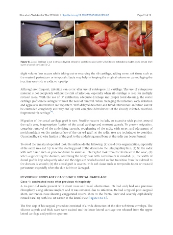
Figure 15. Costal cartilage is cut in straight layered strips(A); septal extension graft with bilateral extended spreader grafts carved from
layers of costal cartilage (B, C)
slight volume loss occurs while taking out or recarving the rib cartilage, adding some soft tissue such as
the mastoid periosteum or temporalis fascia may help in keeping the original volume or camouflaging the
junction area such as radix or supratip.
Although not frequent, infection can occur after use of autologous rib cartilage. The use of autogenous
material is not completely without the risk of infection, especially when rib cartilage is used for multiply
revised cases. With the use of IV antibiotics, adequate drainage and proper local dressing, the costal
cartilage graft can be salvaged without the need of removal. When managing the infection, early detection
and aggressive intervention are important. With delayed detection and timid intervention, infection cannot
be controlled completely and may end up with complete debridement of the already infected, resorbed,
[16]
fragmented rib cartilage .
Migration of the costal cartilage graft is rare. Possible reasons include, an excessive wide pocket around
the radix area, inappropriate fixation of the costal cartilage and remnant capsule. To prevent migration,
complete removal of the underlying capsule, roughening of the radix with rasps, and placement of
perichondrium on the undersurface of the carved graft at the radix area are techniques to consider.
Occasionally, a K-wire fixation of the graft to the underlying nasal bone at the radix can be performed.
To avoid the unnatural operated look, the authors do the following: (1) avoid over-augmentation, especially
at the radix area and try to set the starting point of the dorsum to the interpupillary line; (2) fill the radix
with soft tissue such as perichondrium to avoid an interrupted look from the forehead to the nose; (3)
when augmenting the dorsum, narrowing the bony base with osteotomies is avoided; (4) the width of
dorsal graft is kept adequately wide and the edges are beveled/carved so that transition from the sidewall to
the dorsum is smooth; (5) the dorsal graft is covered with soft tissue such as temporalis fascia or mastoid
periosteum especially when the skin is thin or damaged.
REVISION RHINOPLASTY CASES WITH COSTAL CARTILAGE
Case 1: contracted nose after previous rhinoplasty
A 28-year-old male present with short nose and nasal obstruction. He had only had one previous
rhinoplasty using silicone implant and it was removed due to infection. He had a typical post-surgical
short, contracted nose showing exaggerated nostril show in the frontal view and severely cephalically
rotated nasal tip with low-set nasion in the lateral view [Figure 18A-C].
The first step of his surgical procedure consisted of a wide dissection of the skin-soft tissue envelope. The
silicone capsule and thick scars were excised and the lower lateral cartilage was released from the upper
lateral cartilage and pyriform aperture.