
Page 33 - Read Online
P. 33
Page 14 of 16 Won et al. Plast Aesthet Res 2019;6:6 I http://dx.doi.org/10.20517/2347-9264.2018.82
A B C
D E
F G H
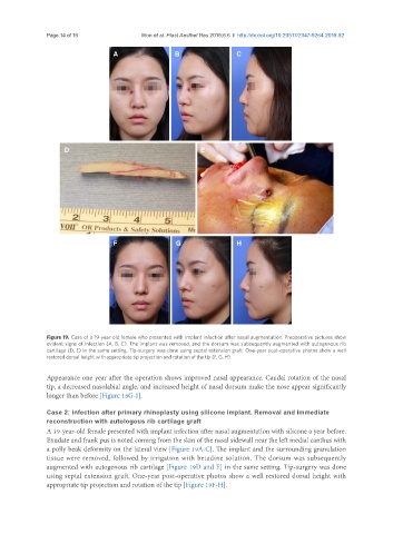
Figure 19. Case of a 19-year old female who presented with implant infection after nasal augmentation. Preoperative pictures show
evident signs of infection (A, B, C). The implant was removed, and the dorsum was subsequently augmented with autogenous rib
cartilage (D, E) in the same setting. Tip-surgery was done using septal extension graft. One-year post-operative photos show a well
restored dorsal height with appropriate tip projection and rotation of the tip (F, G, H)
Appearance one year after the operation shows improved nasal appearance. Caudal rotation of the nasal
tip, a decreased nasolabial angle, and increased height of nasal dorsum make the nose appear significantly
longer than before [Figure 18G-I].
Case 2: infection after primary rhinoplasty using silicone implant. Removal and immediate
reconstruction with autologous rib cartilage graft
A 19-year-old female presented with implant infection after nasal augmentation with silicone a year before.
Exudate and frank pus is noted coming from the skin of the nasal sidewall near the left medial canthus with
a polly beak deformity on the lateral view [Figure 19A-C]. The implant and the surrounding granulation
tissue were removed, followed by irrigation with betadine solution. The dorsum was subsequently
augmented with autogenous rib cartilage [Figure 19D and E] in the same setting. Tip-surgery was done
using septal extension graft. One-year post-operative photos show a well restored dorsal height with
appropriate tip projection and rotation of the tip [Figure 19F-H].