
Page 38 - Read Online
P. 38
Wu et al. Plast Aesthet Res 2019;6:5 I http://dx.doi.org/10.20517/2347-9264.2018.74 Page 3 of 9
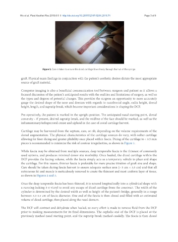
Figure 1. Care is taken to ensure the diced cartilage flows freely through the hub of the syringe
graft. Physical exam findings in conjunction with the patient’s aesthetic desires dictate the most appropriate
source of graft material.
Computer-imaging is also a beneficial communication tool between surgeon and patient as it allows a
focused discussion of the patient’s anticipated results with the realities and limitations of surgery, as well as
the types and degrees of potential changes. This provides the surgeon an opportunity to more accurately
gauge the desired shape of the nose and dorsum with regards to nasofrontal angle, radix height, dorsal
height, length, and supratip break, which become important considerations in shaping the DCF.
Pre-operatively, the patient is marked in the upright position. The anticipated nasal starting point, dorsal
convexity - if present, desired supratip break, and the midline of the face should be marked, as well as the
inframammary/infrapectoral crease and xiphoid in the case of costal cartilage harvest.
Cartilage may be harvested from the septum, ears, or rib, depending on the volume requirements of the
dorsal augmentation. The physical characteristics of the cartilage sources do vary, with softer cartilage
allowing for finer dicing and greater pliability once placed within fascia. Dicing of the cartilage to < 0.5 mm
pieces is recommended to minimize the risk of contour irregularities, as shown in Figure 1.
While fascia may be obtained from multiple sources, deep temporalis fascia is the thinnest of commonly
used options, and produces minimal donor site morbidity. Once healed, the diced cartilage within the
DCF provides the lasting volume, while the fascia simply acts as a temporary vehicle to place and shape
the cartilage. For this reason, thinner fascia is preferable for more precise titration of graft size and shape.
Care should be taken during fascia harvest to ensure adequate surface area (> 5 cm × 3.5 cm) and that all
extraneous fat and muscle is meticulously removed to create the thinnest and most uniform layer of tissue,
as shown in Figures 2 and 3.
Once the deep temporalis fascia has been thinned, it is sutured longitudinally into a cylindrical shape with
a running-locking 5-0 vicryl to avoid any escape of diced cartilage from the construct. The width of the
cylinder is determined by the desired width as well as height of the patient’s bridge, generally in a range
between 3.2-3.5 cm of fascia diameter. One end of the fascia is then closed and filled with an estimated
volume of diced cartilage, then placed along the nasal dorsum.
The DCF will contract and dehydrate when healed, so every effort is made to remove fluid from the DCF
prior to making measurements for its final dimensions. The cephalic end of the DCF is placed at the
previously marked nasal starting point, and the supratip break marked caudally. The fascia is then closed