
Page 24 - Read Online
P. 24
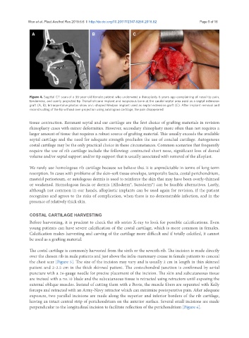
Won et al. Plast Aesthet Res 2019;6:6  I  http://dx.doi.org/10.20517/2347-9264.2018.82                                               Page 5 of 16


                A                            B                   C


               Figure 4. Sagittal CT scan of a 36-year-old female patient who underwent a rhinoplasty 6 years ago complaining of nasal tip pain,
               tenderness, and overly projected tip. Dorsal silicone implant and suspicious bone at the caudal septal area used as a septal extension
               graft (A, B); Intraopertaive photos show an L-shaped Medpor implant used as septal extension graft (C). After implant removal and
               reconstructing of the tip without over projection using autologous cartilage, the pain disappeared


               tissue contraction. Remnant septal and ear cartilage are the first choice of grafting materials in revision
               rhinoplasty cases with minor deformities. However, secondary rhinoplasty more often than not requires a
               larger amount of tissue that requires a robust source of grafting material. This usually exceeds the available
               septal cartilage and the need for adequate strength precludes the use of conchal cartilage. Autogenous
               costal cartilage may be the only practical choice in these circumstances. Common scenarios that frequently
               require the use of rib cartilage include the following: contracted short nose, significant loss of dorsal
               volume and/or septal support and/or tip support that is usually associated with removal of the alloplast.

               We rarely use homologous rib cartilage because we believe that it is unpredictable in terms of long-term
               resorption. In cases with problems of the skin-soft tissue envelope, temporalis fascia, costal perichondrium,
               mastoid periosteum, or autologous dermis is used to reinforce the skin that may have been overly-thinned
               or weakened. Homologous fascia or dermis (Alloderm®, Surederm®) can be feasible alternatives. Lastly,
               although not common in our hands, alloplastic implants can be used again for revision, if the patient
               recognizes and agrees to the risks of complication, when there is no demonstrable infection, and in the
               presence of relatively thick skin.


               COSTAL CARTILAGE HARVESTING
               Before harvesting, it is prudent to check the rib series X-ray to look for possible calcifications. Even
               young patients can have severe calcification of the costal cartilage, which is more common in females.
               Calcification makes harvesting and carving of the cartilage more difficult and if totally calcified, it cannot
               be used as a grafting material.


               The costal cartilage is commonly harvested from the sixth or the seventh rib. The incision is made directly
               over the chosen rib in male patients and just above the infra-mammary crease in female patients to conceal
               the chest scar [Figure 5]. The size of the incision may vary and is usually 2 cm in length in thin skinned
               patient and 2-2.5 cm in the thick skinned patient. The costochondral junction is confirmed by serial
               puncture with a 26-gauge needle for precise placement of the incision. The skin and subcutaneous tissue
               are incised with a no.10 blade and the subcutaneous tissue is retracted using retractors until exposing the
               external oblique muscles. Instead of cutting them with a Bovie, the muscle fibers are separated with Kelly
               forceps and retracted with an Army-Navy retractor which can minimize postoperative pain. After adequate
               exposure, two parallel incisions are made along the superior and inferior borders of the rib cartilage,
               leaving an intact central strip of perichondrium on the anterior surface. Several small incisions are made
               perpendicular to the longitudinal incision to facilitate reflection of the perichondrium [Figure 6].